Download
1 / 44
680 likes | 1.4k Views
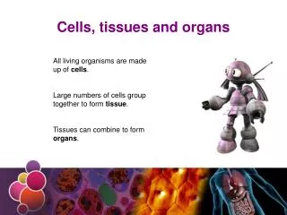
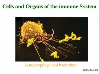
Cells, Tissues, and Organs of the Immune System. Introduction. Knowledge of the structural and ultrastructural details of the immune system is necessary to understand its functions. Distinct compartments that are interconnected by the blood and lymphatic system.

E N D
Introduction • Knowledge of the structural and ultrastructural details of the immune system is necessary to understand its functions. • Distinct compartments that are interconnected by the blood and lymphatic system. • The immune response is coordinated at a system level and complex series of physiologic events interact in vivo to influence the outcome of immune response. • The immune system is integrated with other systems; namely the neural and endocrine systems which can influence the immune response.
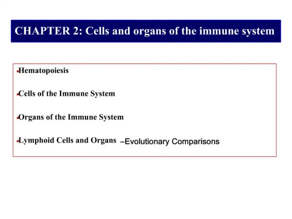
Hematopoiesis • The process of blood cell • Proliferation, • Differentiation, and • Maturation • Sites of hematopoeisis • Bone marrow in adults • Spleen and liver of fetus
Developmental Sites of Hematopoiesis Neonate Fetus Adult Yolk Sac ? Fetal Liver Spleen Bone Marrow
Hematopoietic Tissue • Bones: • Containing hematopoietic marrow (red marrow) • Flat bones of pelvis • Vertebrae • Skull • Ribs and sternum
The Process of Hematopoiesis • Hematopoiesis begins with the stem cell (precursor cells) • Totipotent stem cell • Primitive cell • Potential to turn into any blood cell • Differentiation follows along 2 cell lines (pathways) • Myeloid • Lymphoid
Myeloid Cell Line • Granulocytes (4,000 - 6,000/µl) • Neutrophils 50 - 70% of WBCs • Segments = mature cells • Bands = immature cells • Eosinophils: 1 - 4% • Increase during parasitic infections • Basophils: < 1 % • Involved in allergic reactions
Neutrophil 50-70% of all leukocytes, circulate in the blood, phagocytic, take up neutral dye
Eosinophil 1.5% of leukocytes, release destructive enzymes to destroy invaders, stained with eosin
Basophil Take up basic dye, release histamine, circulate in the blood
Myeloid Cell Line • Monocytes /macrophages • 2-8% of WBCs in blood • Platelets: 150,0000 - 400,000/µl • Red blood cells (RBC): erythrocytes: • 4.2 - 6.2 million/µl
Monocyte 5% of leukocytes, circulates in the blood for a few hours, then crawls into tissues, enlarges and differentiates into...
Macrophage (“big eater”) Phagocytic, very long-lived, some migrate throughout the body, others are resident in tissues (especially lymph nodes)
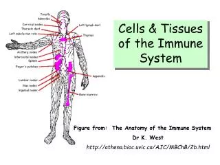
The Lymphatic System Two main functions: 1) Return tissue fluid to circulation 2) Fights infection - both specific and non- specific resistance. Lymph- fluid carried by lymphatic vessels
Lymphocyte Distribution • Lymphocytes lodge in secondary lymphoid organs where they expand clonally upon contact with specific antigens. • Lymphocytes recirculate between secondary organs via blood and lymphatic systems (trafficking). • With the exception of some sites, lymphocytes are widely dispersed in the body.
Lymphoid Cells • Lymphocytes (25-35% of WBCs) • T-cells: 70% of lymphocytes • Cell mediated immunity • B-cells: 20-25% of lymphocytes • Humoral immunity
T Cell T cell B Cell B cells and T cells look alike but have important differences Mature lymphocytes have antigen-specific receptors
Mechanisms of Regulation of Hematopoiesis • Control of cytokine production by stromal cells (altering the microenvironment) • Movement of developing cells from one microenvironment to another • Production of cytokines by non- stromal cells • Up- or down-regulation of cytokine receptor expression by developing cells • Removal of developing (and developed) cells by apoptosis
Critical Cytokines in Hematopoiesis • Interleukin 3 (IL-3) • Produced by T cells (Th1 and Th2) • Binds to IL-3 Receptors on progenitors • Maintains stem cells and early progenitors • Induces proliferation • Does not appear to induce differentiation
Critical Cytokines in Hematopoiesis • Stem Cell Factor • AKA: Steel Factor, Mast Cell Growth Factor, c-kit ligand • Produced by stromal cells • Binds to c-kit on progenitors • Maintains stem cells and early progenitors • Induces proliferation • Does not appear to induce differentiation
Critical Cytokines in Hematopoiesis • Factors important for late progenitors • Erythropoietin (EPO) • needed for red blood cell development • GM-CSF (Colony Stimulating Factor) • works on common granulocyte/ monocyte precursor • G-CSF • works on granulocyte-committed progenitors • M-CSF • works on monocyte /macrophage progenitors
Growth Factor Stromal Cell Stem Cell Response Growth Factor Receptor Stromal Cell Stem Cell No Response Stromal Cell Stem Cell No Response
Bone marrow • The major hematopoietic organ in humans. • Hematopoiesis is facilitated by a mixture of cells and extracellular matrix components. • All blood cell types except mature T cells are found in its cavities. • B cell generation and development occurs in a radial direction towards the center of the bone. • Growth factors, cytokines, and reticular stroma are all important in B cell development.
Thymus • Bilobed organ in the anterior mediastinum. • Grows until puberty then it progressively involutes. • Removal of thymus after birth? • Two types of epithelial cells (endoderm and ectoderm) • Lobes are divided by trabeculae into lobules. • Primary site of T cell development. • Composed of three areas: - Subcapsular zone→ earliest progenitor cell. - Cortex→ Developing T cells undergoing selection. - Medulla→ mature T cells. • >95% of T cell progenitors die in the thymus.
Spleen • Located in left upper abdominal quadrant. • Functions to filter blood from microbes and dead RBCs. • Main site for response to blood-borne antigens and T-independent antigens. • Composed of red pulp (RBCs and macrophages) and white pulp (lymphocytes) • It lodges 25% of the total lymphocytes of the body. • T Cells: Periarteriolar lymphoid sheaths (PALS). • B cells: Primary (resting) and secondary (activated) follicles. • Marginal zones: T cells, B cells, and macrophages.
Lymph Nodes • Bean shaped, usually clustered in groups. • Strategically located throughout the body. • Function to concentrate lymph-borne antigens for presentation to T cells. • Structure: - Cortex (B cells) - Paracortex (T cells) - Medulla( B cells, T cells, and macrophages). • Circulating lymphocytes enter lymph nodes via specialized high endothelial venules (HEVs). • Lymphadenopathy: proliferation in response to infection.
Mucosa-associated lymphoid tissue (MALT) • Respiratory and Gastrointestinal tract(NALT and GALT). • Contain a specialized epithelial cell type (M cell) which engulfs antigens. • Rich in IgA producing plasma cells. • Involved in the establishment of oral tolerance.
Intraepithelial lymphocytes • The mucosa of gastrointestinal, respiratory, and reproductive tracts contain large number of lymphocytes. • >90% T lymphocytes, 50% CD8+ of γδ type. • Develop without the influence of the thymus? • Direct Ag recognition, no need for MHC. • Secrete cytokines that cause immune suppression at the mucosa. • Oral tolerance.
Skin (cutaneous Immune system) • The major physical barrier. • Dendritic cells. • Epidermis has many Langerhan’s cells. • T cells (intraepidermal) mainly CD8+ of γδ type. • Dermis full of macrophages and T cells.
Lymphocyte Recirculation (Trafficking) and Homing • Moving of lymphocytes via blood and lymphatics from one lymphoid tissue to another. • A lymphocyte makes a tour of the body (Blood→ Tissue→ Lymphatic system → Blood) once or twice daily ensuring antigen contact. • Mostly T cells. Naive T cells circulate until they find an Ag or they will die. • B cells have less requirement to recirculate. • Recirculation and homing are regulated by receptor- ligand interactions (selectins, addressins, integrins).
Lymphocyte extravasation • Lymphocyte transport out of the blood stream into tissues. • Four steps: - Primary adhesion to endothelium. - Lymphocyte activation. - Secondary adhesion (arrest). - Transmigration/ chemotaxis.