Download
1 / 43
1.04k likes | 3.53k Views
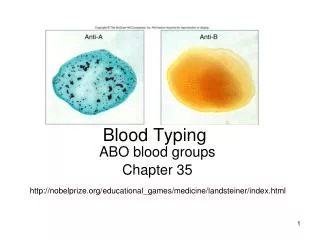
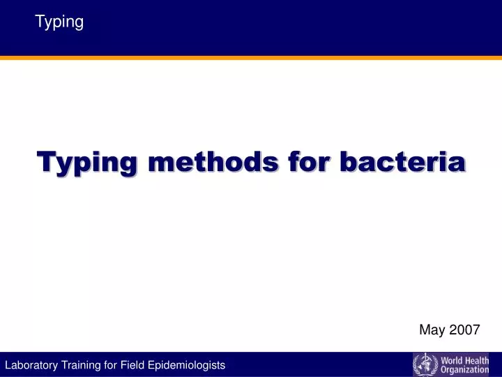
Typing methods for bacteria. Typing. May 2007. Learning objectives. At the end of the presentation, participants should: Identify situations when typing is relevant Know different methods of typing Understand problems that arise when using typing methods. Isolate versus Strain.

E N D
Typing methods for bacteria Typing May 2007
Learning objectives At the end of the presentation, participants should: • Identify situations when typing is relevant • Know different methods of typing • Understand problems that arise when using typing methods
Isolate versus Strain Isolate: a pure culture derived from a single colony that is presumed to arise from a single organism/bacterium Strain: a set of isolates, that when typed are indistinguishable from each other and can be differentiated from other isolates OUTBREAK STRAIN 4 OUTBREAK STRAIN STRAIN 5 STRAIN 2 STRAIN 6 Laboratory Training for Epidemiologists Laboratory Training for Field Epidemiologists STRAIN 3
A simple question ? Are these isolates the same or different? Through a typing method we are looking for: • Epidemiologically linked isolates that represent the clonal expansion of a single precursor • Clonal isolates are the same type and unrelated isolates have a different type Laboratory Training for Epidemiologists Laboratory Training for Field Epidemiologists
Typing system evaluation criteria Laboratory Training for Epidemiologists Laboratory Training for Field Epidemiologists
Phenotype & genotype bacteria Capsule Polysaccharides Fimbirae (M-protein) GENOTYPE (Chromosomal & plasmid DNA) PHENOTYPE Teichoid acid Lipoteichoic acid Different Enzymes Peptidoglycan Laboratory Training for Epidemiologists Laboratory Training for Field Epidemiologists
Typing methods Phenotypic • Rely on expression of phenotypic characteristics (genetically coded) • Antibiotic resistance, antigens etc. Genotypic • Analysis of the genetic material • DNA, RNA Laboratorz Training for Epidemiologists Laboratory Training for Field Epidemiologists
Phenotypic techniques Serotyping Phage typing Antimicrobial resistance monitoring Multilocus enzyme electrophoresis (MLEE) Other: • Protein profiling – SDS PAGE, immunoblotting • Based on nutritional requirement e.g. auxotyping • Biotyping • Bacteriocin typing Laboratory Training for Epidemiologists Laboratory Training for Field Epidemiologists
Phenotypic techniques Serotyping • Antigenetic determinants expressed on the cell surface • Still widely used for Salmonella, Shigella, Neiseria, E. coli, V cholerae • Slide/ tube agglutination • LIMITATION: Requires extensive stock of absorbed/monoclonal sera (e.g. >2200 antisera required for definitive Salmonella typing) Phage typing • Viruses that infect and destroy bacterial cells –Bacteriophage • The resistance or susceptibility of strains is used for differentiation • LIMITATION: Technically demanding, time consuming, typeability is an issue Laboratory Training for Field Epidemiologists
Phenotypic techniques Antibiotic susceptibility testing • Based on susceptibility of bacterial isolates to a panel of antimicrobial agents • Routinely performed on clinical isolates • A reasonable preliminary indicator to initiate epidemiological action LIMITATIONS: • Antibiotic resistance under extraordinary selective pressure • Multiple mechanisms for a strain to become abruptly resistant Laboratory Training for Epidemiologists Laboratory Training for Field Epidemiologists
Phenotypic Techniques Phenotypic characteristics can vary in different conditions • Antibiotic resistance can be expressed under antibiotic pressure Methods are not very discriminatory Laboratory Training for Epidemiologists Laboratory Training for Field Epidemiologists
MLEE Characterizes the cellular proteins by electrophoretically separating them in a gel matrix Exposing the gel to chromogenic substrates (that react with the enzymes) Limitation: Complexity of interpretation Laboratory Training for Field Epidemiologists
Phenotypic typing system characteristics Laboratory Training for Epidemiologists Laboratory Training for Field Epidemiologists
Phenotypic typing during an outbreak Outbreakof Paratyphi B salmonellosis phage type 1var3 France, 1993 Cases September December July August October November
DNA molecule Source: Wikipedia, created by Michael Ströck Laboratory Training for Epidemiologists Laboratory Training for Field Epidemiologists
Genotypic methods Plasmid profiling Restriction enzyme analysis (REA) Restriction fragment length polymorphism (RFLP) Ribotyping Pulse Field Gel Electrophoresis (PFGE) Random Amplified Polymorphic DNA (RAPD) Nucleic acid sequencing Laboratory Training for Epidemiologists Laboratory Training for Field Epidemiologists
The principle Without amplification • Cutting the DNA in pieces • Visualizing the pieces With amplification • Amplifying (using PCR) parts of the DNA • Visualizing the pieces Laboratory Training for Epidemiologists Laboratory Training for Field Epidemiologists
Genotypic typing methods Methods without prior amplification • Isolation of the pathogen • Extraction of the DNA • Cutting the DNA with Enzymes (restriction endonuclease enzymatically cuts/ “digests” DNA at a specific/ “restricted” nucleotide recognition sequence) • Separation of the pieces by size using an electric field (Gel-Electrophoresis) • Visualization Laboratory Training for Epidemiologists Laboratory Training for Field Epidemiologists
Example of molecular typing Gel-Electrophoresis Size of fragments Cutting locations Laboratory Training for Epidemiologists Laboratory Training for Field Epidemiologists
Restriction Enzyme Analysis (REA) • Extraction of plasmid or chromosomal DNA • Digestion of the DNA at particular sites using specific restriction enzymes • Hundreds of DNA fragments of various sizes (0.5-50Kb) separated by gel electrophoresis • LIMITATION: Complex profiles with hundreds of unresolved or overlapping bands Laboratory Training for Epidemiologists Laboratory Training for Field Epidemiologists
Southern blot analysis of RFLP & ribotyping Better analysis of restriction enzyme patterns • Specific parts of the pieces are detected by pieces of DNA as a probe - Southern Blot • Variation in number & size of fragments detected by the markers are referred to as restriction fragment length polymorphism (RFLP) • Ribotyping: when probes mark ribosomal operons LIMITATIONS: technically complex, organisms with single copy of ribosomal operons Laboratory Training for Epidemiologists Laboratory Training for Field Epidemiologists
Southern/Northern blotting "Blot" DNA to membran Separate DNA fragments on the agarose gel Membrane imprinted with DNA bands Add a labelled probe to the membrane Visualization reveals a band where your probe bound to the target sequence Laboratory Training for Epidemiologists Laboratory Training for Field Epidemiologists
Visualization with a marker _I I I_ _I I I_ _I I I_ _I I I_ _I I I_ _I I I_ _I I I_ __ __ __ __ __ _I I I_ _I I I_ _I I I_ _I I I_ _I I I_ Adapted from: http://lifesciences.asu.edu/resources/mamajis/southern/southern.html Laboratory Training for Epidemiologists Laboratory Training for Field Epidemiologists
Pulsed-field gel electrophoresis (PFGE) • Rare cutting enzymes • Alternate current orientations allow separation of large DNA fragments • Highly discriminatory and reproducible; currently the method of choice for typing a range of bacteria LIMITATIONS: time consuming (≥2 days), expensive, specialized equipment Laboratory Training for Epidemiologists Laboratory Training for Field Epidemiologists
Pulsed-field gel electrophoresis (PFGE) RFLPs of MRSA isolates with similar ABT ST profile as determined by PFGE; only isolates B & C are identical RFLPs of VRE isolates as determined by PFGE; all appear identical RFLPs of two strains (B & C) from a patient as determined by PFGE; both different implying mixed infection; lane A is marker Laboratory Training for Epidemiologists Laboratory Training for Field Epidemiologists
Genotypic typing methods Methods with prior amplification • Extraction of the DNA, separation • Target with primer • Amplification of specific region • Separation of amplicons according to size using an electric field (gel-electrophoresis) Laboratory Training for Epidemiologists Laboratory Training for Field Epidemiologists
Molecular typing Gel-Electrophoresis Size of (amplified) fragments Primer-locations Laboratory Training for Epidemiologists Laboratory Training for Field Epidemiologists
Random Amplification of Polymorphic DNA (RAPD ) Uses short primers that find a lot of targets Different size amplicons Products separated by electrophoresis LIMTATIONS: • Identification of suitable primers • Difficult to interpret differences in the intensity of bands • Inefficient reactions • Amplification of cryptic genetic material (prophages, bacteriophages) Laboratory Training for Epidemiologists Laboratory Training for Field Epidemiologists
RAPD-PCR 60 70 80 90 100% 10 Isolates, two clusters (3 isolates each) Laboratory Training for Epidemiologists Laboratory Training for Field Epidemiologists
Nucleic acid sequencing Enumeration of individual nucleotide base pairs Provides highly reliable and objective data suitable for subsequent quantitative analysis Necessary for virus typing LIMITATIONS: • Locus with sufficient sequence variability • Sequencing of a single locus may not be reliable result • Prohibitively expensive for most settings Laboratory Training for Epidemiologists Laboratory Training for Field Epidemiologists
Multi Locus sequence typing (MLST) Targets different DNA pieces and sequences them Compares results with data banks Pro: highly comparable Con: expensive equipment Laboratory Training for Epidemiologists
Genotypic typing system characteristics Laboratory Training for Epidemiologists
Limitations of typing methods • Discriminatory function • Type of material and pathogen • Reproducibility , cost, technique, etc. • No “gold standard” RESULTS OF A TYPING SYSTEM SHOULD BE CONSIDERED RELATIVE TO THE AVAILABLE EPIDEMIOLOGICAL DATA OR TO THE RESULTS OF OTHER SYSTEMS • The technique used needs to be adapted to the question Laboratory Training for Epidemiologists
Llisteriosis outbreaks, France 1999-2000 Cases October November December January February March 1999 2000 Laboratory Training for Epidemiologists
Interpretation of strain typing data Several factors affect interpretation: Natural biologic variation • Epidemiologically related isolates of the same strain demonstrate minor typing differences due to phenotypic variations or actual genotypic alterations, when collected and examined over an extended interval Technical variations • Limited reproducibility and discriminatory power Laboratory Training for Epidemiologists
Interpretation of typing results Genetic relatedness assessed with clinical and epidemiologic relatedness Restrict analysis to discrete set of isolates (≤30) Identify “index isolate” as starting point for analysis that is defined on the basis of: • Epidemiological data (first case in an outbreak) • Clinical data (initial isolate from patient with multiple infections) • Strain typing data (most common strain type in the set) Laboratory Training for Epidemiologists
Interpretation of typing results Multiple isolates representing a single type are most appropriately designated “indistinguishable” No typing method confirms that entire genomes of two organisms are identical Indistinguishable vs. closely related vs. possibly related • Final assessment lies with integration of molecular and epidemiological analyses Laboratory Training for Epidemiologists
Problems with result interpretation Types versus subtypes Isolates assigned as different types if differ in some specified manner (2 or more band shift in S blot) Isolates that differ but not sufficiently to be designated as distinct types are designated as subtypes of similar types Restriction fragments of different sizes may represent same chromosomal DNA Insertion or deletion of extra-chromosomal DNA such as bacteriophage DNA DNA fragment data are not suitable for quantitating genetic relatedness among different isolates Laboratory Training for Epidemiologists
Application of typing systems Detection of outbreaks: Concept of “prior probability”; rigorous epidemiological investigation and data to avoid misleading results Epidemiological investigation • Increased prevalence • Same bacteria species from a cluster of cases • Multiple isolates with distinct biotype/ AST pattern Typing technique with good reproducibility & discriminatory power Request for Molecular typing Laboratory Training for Epidemiologists
Applications of typing systems Distinguish relapse from re-infection Identify types associated with increased transmission & virulence Emergence of new types ; implications on control measures Clonality of acute infection: • Infection vs. colonization vs. sample contamination • Pseudo-outbreaks related to: • Clinician or clinical entity • Laboratory • Case finding • Chance clustering Laboratory Training for Epidemiologists
Phenotypic vs. genotypic typing of Staphylococcus aureus Laboratory Training for Epidemiologists
To summarize Typing data are most appropriately evaluated in the context of a hypothesis and questions thoughtfully developed by the clinician or the epidemiologist. They should augment rather that replace those analyses Typing is performed independently by the laboratory to avoid any bias but the results are considered collaboratively Laboratory Training for Epidemiologists Laboratory Training for Epidemiologists
Typing Developed by the Department of Epidemic and Pandemic Alert and Response of the World Health Organization with assistance from: European Program for Intervention Epidemiology Training Canadian Field Epidemiology Program Thailand Ministry of Health Institut Pasteur