Download
1 / 8
0 likes | 6 Views
Diabetes is a chronic autoimmune disease characterized by the inability of body to produce or respond to insulin a hormone required by body to burn glucose for energy. Type I Diabetes mellitus, also known as Insulin Dependent Diabetes mellitus is a most frequent chronic disease of childhood, afflicts 0.2-0.3% of human individuals due to auto immune destruction of insulin secreting pancreatic u03b2 cells. GAD65 is the major auto antigen in Insulin Dependent Diabetes Mellitus (IIDM). Thus, this project is aimed at expression of GAD65 in E. coli. GAD65 gene was cloned into pET-28a

E N D
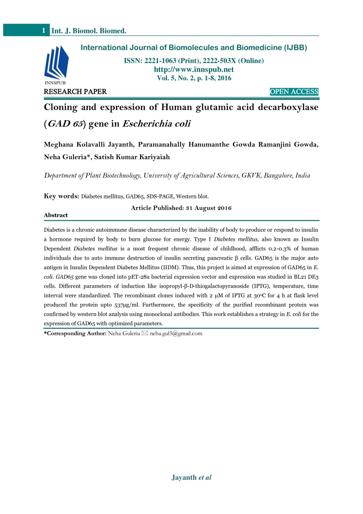
1 Int. J. Biomol. Biomed. International Journal of Biomolecules and Biomedicine (IJBB) ISSN: 2221-1063 (Print), 2222-503X (Online) http://www.innspub.net Vol. 5, No. 2, p. 1-8, 2016 RESEARCH PAPER RESEARCH PAPER OPEN ACCESS OPEN ACCESS Cloning and expression of Human glutamic acid decarboxylase (GAD 65) gene in Escherichia coli Meghana Kolavalli Jayanth, Paramanahally Hanumanthe Gowda Ramanjini Gowda, Neha Guleria*, Satish Kumar Kariyaiah Department of Plant Biotechnology, University of Agricultural Sciences, GKVK, Bangalore, India Key words:Diabetes mellitus, GAD65, SDS-PAGE, Western blot. Article Published: 31 August 2016 Abstract Diabetes is a chronic autoimmune disease characterized by the inability of body to produce or respond to insulin a hormone required by body to burn glucose for energy. Type I Diabetes mellitus, also known as Insulin Dependent Diabetes mellitus is a most frequent chronic disease of childhood, afflicts 0.2-0.3% of human individuals due to auto immune destruction of insulin secreting pancreatic β cells. GAD65 is the major auto antigen in Insulin Dependent Diabetes Mellitus (IIDM). Thus, this project is aimed at expression of GAD65 in E. coli. GAD65 gene was cloned into pET-28a bacterial expression vector and expression was studied in BL21 DE3 cells. Different parameters of induction like isopropyl-β-D-thiogalactopyranoside (IPTG), temperature, time interval were standardized. The recombinant clones induced with 2 μM of IPTG at 30oC for 4 h at flask level produced the protein upto 537μg/ml. Furthermore, the specificity of the purified recombinant protein was confirmed by western blot analysis using monoclonal antibodies. This work establishes a strategy in E. coli for the expression of GAD65 with optimized parameters. *Corresponding Author: Neha Guleria neha.gul5@gmail.com Jayanth et al
2 Int. J. Biomol. Biomed. Introduction One isoform has a molecular size of 65kDa and is Diabetes mellitus is a disease or disorder that is termed GAD 65, while the second one, of 67kDa size, gaining importance and increasing at the alarming is termed GAD67 (Towns and Pietropaolo, 2011). The rate, which is also called as hyperglycemia where the GAD65 enzyme isolation in large quantities from the blood glucose level is higher than the normal. human pancreatic (beta cells) tissue is unrealistic so According to World Health Organization, there are the expression of GAD 65 enzyme as a recombinant mainly two principle types of diabetes; Type 1 protein in heterologous system is mandatory. The diabetes (juvenile or insulin dependent diabetes) and large scale production of GAD 65 enzyme currently type 2 diabetes (insulin independent diabetes).Type involves the use of baculovirus infected Sf9 insect 1diabetes an autoimmune form of diabetic disorder cells and methylo-trophic yeast (Mauch et al., 1993; where the insulin producing β cells of the pancreatic Moody et al., 1995). However, these expression islets are destroyed. It is commonly seen in children system are technically, economically demanding and and the worldwide numbers of prevalent cases of T1D highly vulnerable to contamination. in children (<15 years) have increased and the estimates indicate that there are almost 500,000 Number of heterologous biopharmaceutical proteins expressed in E. coli which are at commercial level are innumerable when compared to all other hosts system this is because of reasons like, it is inexpensive, offers rapid culture times and the ability to achieve high biomass and high protein yields (Assenberg et al., 2013). E. coli expression system remain to be the first choice for laboratory investigations, scaling up activities at commercial level and are useful benchmark for comparison among various expression platforms. The sufficient quantity of soluble and functional recombinant GAD 65 protein is required to carry out any molecular and immunological studies. Poor expression of protein because of cellular toxicity and formation of inclusion bodies are the major hindrance in recombinant protein expression. Therefore, this study was carried out for the optimization of different parameters to enhance GAD 65 protein expression and purification in E. coli. children aged under 15 years with T1D worldwide (Patterson et al., 2014). In this type of diabetes, the autoimmune response occurs against self-antigens which include insulin, intracellular membrane proteins such as GAD, IA2, ZnT8 transporter protein (La Torre and Lernmark, 2010; Wenzlau et al., 2009). There is currently no cure for T1D and the only available treatment is insulin therapy. Insulin therapy can lead to hypoglycemia if the doses are not taken properly without monitoring blood glucose level and patients requires two or more injections of insulin daily which is painful. (Alvarez et al., 2013).In this regard, GAD65 a major auto antigen have much potential as an important marker for the prediction, diagnosis and in the prevention of T1D in humans. Autoantibodies to GAD65 are observed months to years before the clinical onset of diabetes and are present in the sera of 70–80% of patients with T1D Materials and methods and this anti-GAD 65 antibodies are now serving as Bacterial strains, plasmids and growth conditions an important marker for the prediction and diagnosis The E. colistrains DH5α and BL21 (DE3) were used of type 1 diabetes (Jayakrishnan et al., 2011; Oak et for the cloning and expression experiments. The al., 2011; Wang et al., 2012). expression vector pET-28a used in this study was Glutamic acid decarboxylase is an enzyme which is purchased from Novogen, USA. The pTNT vector carrying GAD 65 gene was provided by Ake involved in the production of Gamma amino butyric Lernmark, Professor, Lund University Sweden. The E. acid (GABA) by the decarboxylation of glutamate is coli was grown in Luria Bertini Broth (LB) at 37°C an inhibitory neurotransmitter in neurons and with shaking 200rpm or on LB plates solidified with pancreatic beta cells. This GAD exists in two major 1.5 w/v agar. protein isoforms. Jayanth et al
3 Int. J. Biomol. Biomed. As per the requirement LB broth and agar plates were followed by a thrombin protease cleavage site. Two supplemented with kanamycin in a final stop codons are included in the vector at the C- concentration of 50 mg/L. The GAD 65 gene was terminal cloning site with kanamycin resistance cloned and maintained in pTNT cloning vector. (nptII) gene as selection marker and poly linker Bacterial expression vector pET-28a was selected to sequence with multiple cloning sites (Fig. 1.). This express the gene encoding for GAD 65 protein in E. vector replicates in E. coli through it sp BR322 origin coli. It contains T7 promoter driven expression of of replication. Plasmid DNA was isolated and purified recombinant proteins with the addition of a 19 amino using QIA prep Miniprep Kit (Qiagen, USA) according acid N-terminal fusion tag containing a 6X His tag to the manufacturer protocol. Fig. 1.Vector map of recombinant pET-28a_GAD 65. Primer designing for the cloning of GAD65 gene Oligonucleotides primers were designed for PCR amplification of GAD65 gene. To facilitate the cloning of GAD 65 gene into the expression vector pET-28a, NcoI restriction site was inserted in the forward primer (5’-atatcatg CCATGGCTAGCCCAGGCT-3’) and XhoI site in the reverse primer sequence (5’- ccg CTCGAGTAAATCTTGTCCAAGGCG-3’) due to the absence of the two sites in the GAD 65 gene. Cloning of GAD65 gene into bacterial expression vector pET-28a The GAD 65gene in the pTNT vector was used as a template and was amplified meraseusing a pair of gene specific primers GAD65 XhoI forward primer and GAD65 NcoI reverse primer in 50 µL reaction mixture. The programme is as follows; Initial denaturation at 98°C for 3 minutes followed by 30 cycles of final denaturation at 98°C for 10 sec, annealing at 60°C for 30 sec and extension at 68°C for 1 min with a final extension for 7 min at 68°C. The PCR product was resolved on 1 per cent agarose gel and the 1.75 Kb GAD 65gene was eluted from the agarose using Qiagen gel elution kit. The purified PCR product (GAD 65 gene) was restriction digested in 50µL reaction mixture (2µg DNA, XhoI and NcoI). The bacterial expression vector pET-28a was restriction digested with XhoI and NcoI. The pET-28avector carries with Pfu poly an N-terminal 6X His tag. The GAD65 gene was cloned to the upstream of N-terminal coding portion of pET- 28a vector (Fig. 1.). Jayanth et al
4 Int. J. Biomol. Biomed. The digested PCR product was ligated into digested pET- 28a vector in 1:3 ratio using T4 DNA ligase enzyme. The ligated mixture was transformed into E. coliDH5α competent cells by electroporation as described by Sam brook and Russel (2001). Transform ants were screened using 50 mg/l kanamycin selection. The column was washed with 1X PBS to remove unbound foreign or host proteins. Then the GAD 65 protein was eluted from the column using different concentration of imidazole (50, 150,250 mM). The concentration of purified GAD 65 protein isolated from E. coli BL 21 cell was measured by using To confirm the recombinant clones, the colony PCR Bradford protein assay (Bradford, 1976). was done using a pair of gene specific primers. SDS PAGE and Western blot analysis Further these clones were also confirmed by The purified GAD 65 protein was analyzed by SDS- restriction digestion with NcoI and XhoI and PAGE as per the standard procedure of Lamelli confirmed on 0.8% agarose gel. (1970). The protein samples were prepared by GAD 65 protein expression denaturing the proteins by boiling for 15 min with The E. coli BL 21 competent cells were transformed SDS loading dye. The protein samples were run on 12 with confirmed recombinant clone as described by per cent acrylamide gel along with standard protein Sam brook and Russel (2001) and screened using 50 marker. Further the expression of the GAD 65 protein mg/Lkanamycin selection. The single colony was was confirmed by western blot as per the standard cultured by inoculating it in 10 mL LB broth protocol of Tow bin et al., 1979. The protein bands containing kanamycin (50 mg/L) and incubated at were transferred from SDS-PAGE gel to activated 37°C overnight. From the overnight grown culture PVDF membrane by electro blotting. The unspecific one mL was inoculated into 100 mL LB broth to make protein binding sites were blocked with blocking 1:100 dilution and incubated at 37°C until the OD buffer containing skimmed milk and incubated at reached 0.4-0.6. At this point, different parameters 37°C for 1 h. Then the membrane was washed with were used to optimize GAD 65 protein expression in PBST and incubated for 1 h at 37°C with GAD65 E. coli i.e., different IPTG concentration (0.1, 0.3, 0.5, monoclonal antibody. The binding of the antibody 0.7, 1, 1.5, 2.0, 2.5, 3.0 3.5 and 4.0 µM), temperature was visualized using goat antibody antimouse Ig G (25°C and 30°C) and time interval (4, 8, 12, 16 and 18 conjugate with peroxidase. h). The cells were harvested by centrifugation at 10000 rpm for 15 min at 4°C. Results and discussion GAD 65 protein extraction and purification The harvested cells were suspended in lysis buffer [(1X PBS,), 7µL PMSF and 1mg/m Llysozyme] and incubated on ice for 15 min. Cells were lysed by sonication at 200-300 W for 15 min with alternative 10 sec sonication and 10 sec rest on ice. The cells were sonicated and pelleted out at 10000 rpm for 10 min at 4°C. The supernatant was collected and subjected for purification using Ni-NTA agarose column. The column of Ni-NTA agarose was prepared, equilibrated and activated with 1X PBS. The column was loaded with protein sample and flow throw was collected. Generation 28a_GAD65 The E. colistrain DH5αwas transformed with the ligation mixture and the recombinants were grown on kanamycin selection media. The colony PCR resulted in the amplification of GAD65 gene, amplicon size ~1.75Kb. (Fig. 2.). Further, restriction digestion of recombinant clones with XhoI and NcoI restriction enzymes resulted in linearization of pET-28a vector (~5.23 kb) and the release of the GAD 65 gene (~1.75Kb). This confirms the proper integration of the gene into the expression vector. Thereafter, clones are designated as recombinant clones (Fig. 3.). of recombinant clones of pET- Jayanth et al
5 Int. J. Biomol. Biomed. Fig. 2. Colony PCR of transformed colonies of pET-28 a_GAD 65 gene. M: 1 Kb DNA ladder; lane 1: Negative control, lane 2: Positive control; lane 3-10: PCR amplicon of GAD 65 gene. The optimum growth and maximum expression i.e. 530μg/mL of protein was seen at 2.0 µM IPTG at 30°C after 4 h of induction (Fig. 4.). The decrease in protein expression after 2 µM IPTG concentration is may be due to the fact that further increase in IPTG concentration acts as inhibitory for the bacterial cells. Lower IPTG concentration, lower temperature with longer induction time leads to the slower expression rate of the protein which allows better folding of the protein but reduces yield. The higher IPTG Fig. 3. Confirmation of recombinant pET-28a_GAD concentration with shorter induction time at low temperature may allow higher properly folded protein 65 gene construct with XhoI and NcoI restriction expression which leads to increased yields of protein digestion. M: 1Kb DNA ladder; Lane 1: undigested (Tolia and Joshua- Tor, 2006). As the GAD 65 gene pET-28a vector; lane 2: undigested recombinant pET- was cloned into pET-28a expression vector, it has lacI 28a GAD65 gene construct; lane 3-4: Restriction promoter which codes for repressor protein and a T7 digestedre combinant pET-28a GAD65 construct. promoter which is specific to only T7 RNA Effect of IPTG concentration on gene expression Different parameters like IPTG concentration, induction temperature and length of induction influences both the solubility and yield of the protein. For the expression vector pET-28a_GAD 65, the foreign protein expression was triggered by adding IPTG into the culture medium. The Fig. 4. showed overall growth pattern of recombinant clones on different concentrations of IPTG ranging from 0.1, 0.3, 0.5, 0.7, 1.0, 1.5, 2.0, 2.5, 3.0, 3.5, 4.0 µM. Although the maximum cell growth was observed at 0.3 to 0.5µ M IPTG. polymerase. The control of pET expression system is accomplished through the lac promoter and operator. Addition of IPTG displaces the repressor from the lac operator and induces the expression of T7 RNA polymerase which in turn transcribes the target gene cloned into the pET-28a bacterial expression vector system. By adjusting the concentration of IPTG, expression can be regulated from very low level expression up to the robust, fully induced expression levels commonly associated with pET vectors (German and Eduardo, 2014). Jayanth et al
6 Int. J. Biomol. Biomed. Fig. 4. Effect of IPTG concentrations on bacterial cell growth and GAD 65 protein expression. Effect of different temperature and time interval on gene expression The optimum temperature for E. coli growth is 37°C but induction for recombinant protein expression are carried out at low temperature to retain the solubility (folding) of the expressed protein. The recombinant clones showed maximum growth at 37°C as compared with the other temperatures. Although the cells grew well at 37°C but the protein expression was not maximum at this temperature. At 25°C with 4, 8, 12, Fig. 5. Effect of different induction temperatures and 16, 18 h time interval the protein expression was 259, time intervals on GAD 65 protein expression. The 391, 498, 250 and 245μg/mL respectively while at error bars indicate the standard deviation. 30°C with same length of induction period, the GAD Expression and Purification of GAD 65 protein 65 protein expression was 537, 436, 319, 298 and 256 μg/mL respectively. Hence, E. coli cells induced at After the optimization of the different parameters, the 30°C for 4 h and 25°C for 12 h at 2 μM IPTG histidine tag fused GAD 65 protein expressed in E. concentration showed the highest GAD65 protein coli BL 21 cell was extracted and purified by metal expression of 537 μg/mL and 498 μg/mL respectively affinity chromatography. The purification of protein (Fig. 5.). This may be due to slower expression rate at is essential for any biochemical analysis, therapeutics 25°C when compared to 30°C. The temperature is or structural studies. The GAD 65 protein samples known to influence the expression of recombinant were passed through Ni-NTA agarose column and the proteins in E. coli. When the temperature decreases, optimum elution of GAD65 protein was obtained induction time increases because of reducing culture when 250 mM imidazole was used. Similar result was temperature usually leads to slower growth of reported by Papouchado et al., (1997) where they bacteria and slower rate of protein production (Gong expressed GAD 65 protein as a fusion protein with et al., 2009). The bacterial growth at 37°C causes thioredoxin which helped in the purification of GAD some proteins to accumulate as inclusion bodies, 65 protein. The purified GAD 65 protein expressed in while incubation at 30°C leads to soluble, active E. coli was confirmed by SDS-PAGE where 65 kDa protein (Vera et al., 2007). GAD 65 protein band was observed (Fig. 6A.). Jayanth et al
7 Int. J. Biomol. Biomed. Similarly, Rohmah et al., (2013) reported 65 kDa GAD 65 protein band on SDS-PAGE. This may be due to the correct expression and proper folding of GAD65 protein in E coli. Further it was confirmed by western blotting using monoclonal antibody. The presence of single 65 kDA protein band on the PVDF membrane confirmed the expression of GAD 65 protein (Fig.6B.). A powerful purification method involves the use of peptide affinity tags, which are fused to the protein of interest and used to expedite protein purification via affinity chromatography (Bornhorst and Falke, 2000). Hence, immobilized metal-affinity chromatography (IMAC) can be used to purify recombinant proteins containing a short affinity tag This is in line with Papouchado et al., (1997) who expressed GAD 65 protein in E. coli. The expression and subsequent purification of recombinant proteins are widely employed in biochemical and molecular studies. consisting of polyhistidine residues, theoredoxin residues etc., and the Ni NTA passed protein could keep its function. Fig. 6. SDS-PAGE and western blot analysis of recombinant GAD 65 protein expressed in E. coli. (A) Lane M: Protein ladder; lane 1: Purified protein sample (B) Western blot analysis. Lane M: Protein ladder; lane 1-4: GAD 65 protein samples expressed in E. coli. In conclusion, the different parameters were standardized for the expression of GAD 65 protein in E. coli. The protein produced can be aimed at developing a diagnostic kit for the prediction of insulin-dependent diabetes mellitus (Type-1). Assenberg R, Wan PT, Geisse S, Mayr LM. 2013. Advances in recombinant protein expression for use in pharmaceutical research. Current Opinion in Structural Biology 23, 393-402. Acknowledgements Authors are highly thankful to the Dr. Ake Lernmark, Sweden for providing the GAD 65 gene construct. Bornhorst JA, Falke JJ. 2000. Purification of proteins using polyhistidine affinity tags. Methods Enzymology 326, 245-254. References Alvarez ML, Done SC, Cardineau GA. 2013. Reverse vaccination and treatment of type 1 diabetes using plant produced auto antigens and anti- inflammatory cytokines. Journal of Clinical and Cell Immunology S2:007. DOI: 10.4172/2155-9899.S2-007. Bradford MM. 1976. A rapid and sensitive method for quantitation of microgram quantities of protein utilizing the principle of protein-dye binding. AnalyticalBiochemistry72, 248-254. Jayanth et al
8 Int. J. Biomol. Biomed. German LR, Eduardo AC. 2014.Recombinant protein expression in Escherichia coli: advances and challenges. Frontiers in microbiology DOI: 10.3389/fmicb.2014.00172. Patterson C, Guariguata L, Dahlquist G, Soltesz G, Ogle G, Silink M. 2014. Diabetes in the young- A global view and worldwide estimates of numbers of children with type 1 diabetes. Diabetes Gong Z, LongX, Pan L, Le Y, Liu Q, Wang S, Guo J, Xiao B, Zhou M, Mei D. 2009. Cloning, expression, purification and characterization of the cholera toxin B subunit and triple glutamic acid decarboxylase epitopes fusion protein in Escherichia coli. Protein Expression and Purification 66, 191-197. Research and Clinical Practice 103, 161-175. Rohmah RN, Widyasari S, Aulanni A, Fatchiyah M. 2013. Cloning and expression of GAD 65gene in E. Coli BL 21. Indonesian Journal of Biotechnology 18, 52-57. Jayakrishnan B, Hoke DE, Langendorf CG, Buckle AM, Rowley MJ. 2011. An analysis of the cross-reactivity of autoantibodies to GAD 65 and GAD 67 in diabetes. PLoS ONE 6,1-6. Sambrook J, Russel DW. 2001. Molecular Cloning: A Laboratory Manual, 3rd edn. Cold Spring Harbor, N.Y: Cold Spring Harbor Laboratory Press 576-585. La Torre D, Lernmark A. 2010. Immunology of beta-cell destruction. Advances inExperimental Medicine and Biology 654, 537-83. Tolia NH, Joshua-Tor L. 2006. Strategies for protein co expression in Escherichia coli.Nature method 3, 55-63. Mauch L, Seissler J, Haubruck H, Cook NJ, Abney CC, Berthold Liedvogel B, Scherbaum WA, Northemaim W. 1993. Baculovirus-mediated expression of human 65 kDa and 67 kDa glutamic acid decarboxylases in SF9 insect cells and their relevance in diagnosis of insulin dependent diabetes mellitus. Journal of Biochemistry 699, 704-727. H, Wirbelauer C, Towbin H, Staehelin T, Gordon J. 1979. Electrophoretic transfer of proteins from polyacrylamide gels to nitrocellulose sheets: Procedure and some applications. Proceedings of National Academy of Sciences USA 76, 4350-4354. Vera A, Gonzalez-Montalban N, Aris A, Villaverde Moody AJ, Hejnaes, KR, Marshall MO, Larsen FS, Boel E, Svendsen I, Mortensen E, Dyrberg T. 1995. Isolation by immunologically and enzymatically active human islet glutamic acid decarboxylase 65 overexpressed in Sf9 insect cells. Diabetologia 38, 14-23. A. 2007. The conformationalqualityofinsoluble recombinantproteinsisenhancedat low growth anion-exchange of temperatures. Biotechnology Bioengineering96, 1101- 1106. Wang X, Zhang A, Liu Y, Chen S, Feng Z, Oak S, Radtke J, Torn C, Olsson ML, Hampe CS. 2011. Immunoglobulin subclass profiles of antiidiotypic antibodies to GAD65Ab differ between type 1 diabetes patients and healthy individuals. Scandinavian Journal of Immunology 74, 363-367. Shang W, Maziarz M, Radtke J, Hampe CS. 2012. Anti-Idiotypic Antibody Specificto GAD 65 autoantibody prevents type 1 diabetes in the NOD mouse. PLoS ONE 7, 1-8. Papouchado ML, Valdez SN, Ghiringhelli D, Poskus E, Ermacora, MR. 1997. Expression of properly folded human glutamate decarboxylase 65 as a fusion protein in Escherichia coli. Europian Journal of Biochemistry 246, 350-359. Wenzlau JM, Frisch LM, Gardner, T. J., Sarkar, S., Hutton, JC, Davidson H. 2009, Novel antigens in type 1 diabetes: The importance of ZnT8. Current Diabetes Reports 9, 105-112. Jayanth et al