Download
1 / 16
160 likes | 355 Views
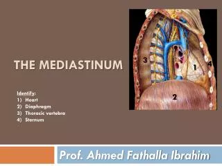
Posterior Mediastinum II. Thoracic Duct. It is the largest lymphatic channel in the body. It conveys most lymph of the body to the venous system(all but the right upper quadrant). Course.

E N D
Thoracic Duct • It is the largest lymphatic channel in the body. It conveys most lymph of the body to the venous system(all but the right upper quadrant).
Course • The Thoracic duct begins below in the abdomen as a dilated sac, the cisterna chyli. It ascends through the aortic opening in the diaphragm, on the right side of descending aorta. It gradually crosses the median plane behind the esophagus and reaches the left border of esophagus at the angle of louis. It then runs upward along the left edge of esophagus to enter the root of neck.
Continued • In the posterior mediastinum, its relations are as follows • Anteriorly, esophagus • Posteriorly, Vertebral bodies • On the left, thoracic aorta • On the right, azygous veins
In the root of the neck, it bends laterally behind the carotid sheath and in front of vertebral vessels. It turns downward in front of phrenic nerve and crosses the subclavian artery to enter the left brachiocephalic vein. • At the root of neck, it receives left jugular, subclavian,braciomediastinal trunks. It conveys lymph to venous system from lower limbs,pelvic cavity, abdominal cavity, left side of thorax and left side of head,neck and left arm.
Azygous Venous System • The Azygous system of veins, on each side of the vertebral column, drains the back and thoraco-abdominal walls, as well as the mediastinal viscera.
Azygous Vein • It forms a collateral pathway between the SVC and IVC and drains blood from the posterior walls of the thorax and abdomen. It is formed by union of the right ascending lumbar vein and the right subcostal vein. • It ascends through the aortic opening in the diaphragm on the right side of the aorta to the level of T5. Here, it arches above the root of right lung to empty into Superior Vena Cava.
Tributaries • Eight lower right intercostal veins • Right Superior Intercostal Vein • Superior and Inferior Hemiazygous vein • Mediastinal veins
Hemi-azygous vein/Inferior Hemi-azygous • It is formed by union of left subcostal and ascending lumbar veins. It ascends on the left side of vertebral column, posterior to the thoracic aorta as far as T9 vertebra. Here it crosses to the right, posterior to aorta,thoracic duct and esophagus, to join the azygous vein. It receives tributaries from Left lower intercostal veins and mediastinal veins.
Superior/Accessory Hemi-Azgous Vein • It is formed by union of fourth to eights intercostal veins. It crosses over T7 or T8 vertebra, posterior to the thoracic aorta and thoracic duct, where it joins the Azygous Vein.