Download
1 / 46
470 likes | 611 Views
SIDs, AW disorders, Asthma, & Plural Disorders Chapters: 31,32,33 & 43. Walsh E. Chavez RRT-NPS. Definition of SIDS. Sudden and unexpected death of an infant for which sufficient cause cannot be found by a death scene investigation, review of the history, and a postmortem

E N D
SIDs, AW disorders, Asthma, & Plural DisordersChapters: 31,32,33 & 43 Walsh E. Chavez RRT-NPS
Definition of SIDS • Sudden and unexpected death of an infant for which sufficient cause cannot be found by a death scene investigation, review of the history, and a postmortem • 1week-1 year old (2-4 months < 6months) • ALTE (Apparent life-threatening event) • Color change • Hypotonia • 70% found in the morning…..
Risk Factors • Lending for intervention • Prone positioning • Maternal smoking • Bottle-feeding vs Breast feeding????
Normal Control of Heart Rate and Breathing • Breathing • Brainstem • Heart rate • Autonomic nervous system
SIDS • Etiology: • Brainstem: controls breathing(Prematurity?) • Central Apnea & periodic breathing • Tests for assessing risk factors • NO test that predicts risk for SIDS • Polysomnography/ pneumogram • Frequency of apnea
Polysomnography • Parameters: Pg. 547 Fig 31-3 • EEG (Brain activity) • EOG (Bilateral Eye Movement) • EMG (Muscle tone) impendence belt -EtcO2 -POX/ TCOM -ECG -Ph Probe graph Figure 31-2 (pg 546) • LaboratorySupervision: PediatricianWho Scores? • Setting: Pediatric Staged with Parent near • Personnel: knowledge of the child's behavior developmental stages
Polysomnography (cont.) • Normal sleep development: • REM and NON-REM • Apnea (3 categories) • Rapid base line • Central: (B’sD’s andColor change) 10-25% • Obstruction:hypopnea & apnea • OB lasting >10sec or 6-8 sec (UAW OB) • Complications: Desat/ > CO2;Day time Sleepiness, behavior changes, cor pulmonale • Treatment: • Removal of adenoidal & tonsils • NCPAP • NICU: Reflux, PofA, Hypoxia, anemia, IVH, Seditaives, seizures, incoordiation with feedings
Home Cardiorespiratory Monitors= Apnea Monitors Alert the caregiver to a cardiorespiratory abnormality Diagnostic devices Training Social/parental implications
Chapter 32 Pediatric Airway Disorders and Parenchymal Lung Diseases
Pediatric Airway Upper airway (Pg. 555 fig. 32-1) Above C3-4 Peds non expandable Cricoid rings(Narrowest portion) -obligate nose breathers 3-6 months Poor coronation between rr& oropharngeal motor skills & large tongues Developement up to 8 years of age • Lower airway: • Trachea > Subdivisions 17-16: adults23 generations…..RSV ? • Airway obstruction: • AP/ Lateral Neck-XRAY • Assessments: RDS, Auscultate UAW: Air movement muffled/stridor
Upper Airway Disorderslesions, inflammatory, abnormal tissue, tone • Supralaryngeal obstruction • Choanal atresia • Pierre Robin syndrome • Deep neck infections • Tonsillar enlargement/Peritonsillar abscess • Obstructive apnea
Upper Airway Disorders (cont.) • Periglottic obstruction (around the glottis) • Epiglottis • Laryngotracheobronchitis (LTB-CROUP) • Table 32-1 Differential Diagnosis • Pg. 559
Epiglottitis Life Threatening =Bacterial infection (H influenza B) • Incidence and etiology • <6 years old • Noninfectious: aspiration of hot liquid, traumatic intubations, blind finger sweep • Signs and symptoms: ABRUPT • Fever, soar throat, dysphagia(drooling) • MUFFLED, retractions, RDS, upright sniffing position • Diagnosis: lateral NECk (Thumb sign) C&S, ABG • Treatment: (small?) ETT, RSI, ceftriaxone, Extubate leak?Bronchoscope, antibotcs
LaryngotracheobronchitisLTB-CROUP(6 months-6 years) • Incidence and etiology: • Parainfluenza Virus 1 • Over several days • Signs and symptoms: • Low grade fever, malasise, rhinorrhea, hoarse voice BARKY COUGH • Diagnosis: • Lateral neck-xray: Steeple sign (SUBglottic) • Treatment: Racemic Epi, hydration, temp ……………..control, humidification, Mist tents, O2, >0.35Fio2= impending resp Failure Intubations
Lower Airway Disorders • Obstruction of the trachea and major bronchi • Tracheomalacia • Congenital tracheal or bronchial stenosis
Lower Airway Disorders pg563 • Foreign body aspiration • Incidence: • Leading cause of accidental death • Signs and symptoms: • Degree of AW OB • Unilateral Wheezing/ reoccurring PNA • Diagnosis: AP CXR/neck and lateral • Laryngeal level • Hyperinflation/Ball valve effect • Bronchoscopy • Treatment: • Removal of object continue to monitor patient
Lower Airway Disorders • Atelectasis • Etiology and pathophysiology: • Failure to reinflate • Signs and symptoms: • Dyspnea • (Severe) V/Q mismatch • RDS • Diagnosis: CXR, B/S, tracheal deviation • Treatment: Hyperinflation therapy, Bronchial hygiene therapy,
Lower Airway Disorders • Bronchiectasis: • Irreversible dilation of the bronical tree • Etiology and pathology • CF/ frequent respiratory infections • Left lower lobes mostly involved • Signs and symptoms: • Chronic cough w/ copious amts of purulent sputum • Diagnosis: CXR and CT • Treatment: CPT, hydration, antibotics,
Lower Airway Disorders • Acute bronchiolitis • Etiology: Viral Respiratory Tract infection related to RSV • Incidence:<1 year of age with BPD, CF, PPHN & CHD • Signs and symptoms: coryza, cough, RDS, wheezing (APNEA), dehydration • Diagnosis:Nasal Swab: +RSV • Treatment:O2, (MV), IV, Supportive care • Prognosis: GOOD
Pneumonia • Viral • 1st:Respiratory syncytial virus (RSV) • Coryza, NASAL CONGESTION, cough & fever • Supportive Care vs. Ribavirin • 2nd:Parainfluenza virus Types1(LTB), 2, and 3(children<5yr) • O2 and supportive care • Influenza virus: • Winter seasons • Vaccinations yearly • Adenovirus: • High rates of M & M. • Overwhelming sepsis • Supportive care
Pneumonia • Bacterial • Incidence: Compromised immune function, recurrent aspiration, malnutrition, daycare, passive cigarette • Etiology: microorganisms colonize in the URT • Signs and symptoms: same as viral • Diagnosis: CXR, > total band count>1500, CRP, Blood Culture • Treatment: oral/ IV antibiotics • 7-14 days • Pox & ABG
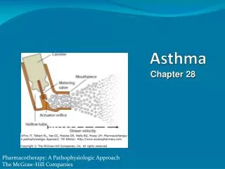
Chapter 33 PG. 582-597 Asthma Most common chronic childhood disease
Pathogenesis • Definition:Chronic inflammatory disorder of the AWs • Mast cells, eosinophils, T lympocytes, IgE, Macrophages, neutrolphils, & epithelial cells • Wheezing &Breathlessness
Pathogenesis of Asthma • Pathophysiology • Chronic airway inflammation • Bronchial hyperresponsiveness • Hypersecretion of mucus • Components of Asthma: • Inflammation -A remodeling • Bronchial constriction -Mucous plugging • AW edema -AW Hyperreponsiveness • RESULTS: • HYPERinflation, atelectasis, hypoxia, V/Q mismatch, Hypercarbia
Risk Factors for Developing Asthma • Allergic response: • IgE • Reversible vs. irreversible • AW remoding=fibrosis • Environmental triggers • Intervention • Remediate & eliminate • Tobacco • Cockroaches/ Dust mites • Molds
National Asthma Education and Prevention Program • Purpose: • to serve as a comprehensive tool to diagnosis and manage asthma • Goals: Box 33-1: • Prevent: Chronic asthma, recurrent • Maintain: NL ADLs, NL/near NL pulmonary function • Optimal pharmacology • Meet family & patient’s expectations
Diagnosis • Medical history: • Symptoms & triggers • Physical examination: • History a stronger factor • Prolongs expiratory phase • RDS, Hyperexpansion • Pulmonary function testing: • FVC, FEV1 <80% predicted, FEV1/FV(< 65% predicted), FEF25-75% • Bronchoprovocational challenges (methacholine 20% decrease=+test) • Exercise: running/biking • Less sensitive HR > 170
Management • Pharmacologic therapy • Long-term control medications • Taken daily • Antiinflammatoryagents/ corticosteroids • Long acting B2 agonist(LABAs) • Salmeterol & Formoterol 30-90 minutes • Methylxanthines • Leukotriene modifiers: inhibits • Cromolyn sodium (Stablizes mast cells) • Immnuomodulators: Binds to IgE
Management • Quick-relief medications: (5-15 minutes) • Short acting B-agonist last 4-6 hours • “rescue” • Anticholenergics • Delivery systems • MDI (Spacers & holding chambers) • DPI • SVN
Management (cont.) • Control of triggers • Identification of allergens • Avoidance and control measures • Immunotherapy • controversial
Management (cont.) • Peak flow monitoring • Peak flow meter • Peak flow diary • Personal best reading • Peak flow zone system • Pg. 593
Patient and Family Education Asthma disease process Medication skills Identification and control of triggers Self-monitoring techniques
Managing Asthma • Exacerbations in the ED • Assessment: PF of airflow, POX • Beta-2 agonists • Corticosteroids
Managing Asthma (cont.) • Hospitalization and respiratory failure BOX 33-8 (Criteria for hospitalization) • Intubation • Elective • Respiratory Fatigue • Mechanical ventilation • Low Vt • PS ventilation Based on the degree of sedation • I:E ratio for adequate ventilation • Prone to pneumothorases, barotrauma, per > PIP, & hypotension
Asthma • Exercise-induced bronchospasm (EIB) • 5-10 minutes after activity • Asthma at school • Teachers • Self esteem • Asthma camps • AHA • SCAMP CAMP
Chapter 43 Pg. 706-714 Disorders of the Pleura
Pleural Effusion • Clinical signs: • Decrease in B/S, Dullness to percussion compared to the contralateral • Tachypnea and pain • Diagnosis: CXR • Tx:Thoracentesis • Laboratory studies: Transudate vs. Exudate, empyema (-Tube) • Causes:BOX 43-2 & 43-3 (pg. 708) • Complications: • pneumothorax • Hemorrhage • Infections
Pneumothorax • Clinical signs: • Chest pain & SOB • Decrease breathsounds on affected side with hyperresonnance • Diagnosis: CXR • Treatment: • Nitrogen washout • Needle aspiration • CT
Thoracostomy Drainage • Indications: Drainge of air/ fluid • Procedure for placement • Conscious sedation • Role of the respiratory therapist • Assist/ Place • Drainages system (3 bottles)
Surgery in the Pleural Space Treatment of empyema Thoracoscopy Chemical pleurodesis