Download
1 / 29
290 likes | 457 Views
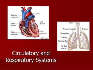
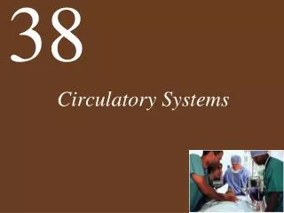
50. Circulatory Systems. A circulatory system consists of: A muscular pump – t he heart A fluid – blood A series of conduits – blood vessels Together these are called the cardiovascular system. Capillary bed injected with dye. www.olympusmicro.com/galleries/abramowitz/images/.

E N D
50 Circulatory Systems
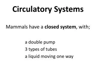
A circulatory system consists of: • A muscular pump – the heart • A fluid –blood • A series of conduits –blood vessels • Together these are called the cardiovascular system Capillary bed injected with dye www.olympusmicro.com/galleries/abramowitz/images/
Some animals do not need circulatory systems: • Single-celled organisms exchange nutrients, gases, wastes directly with their environment • Structures and flattened body shapes increase surface area to enhance exchange between cells and environment • E.g. gastrovascular cavities – highly branched cavity in flatworms and cnidarians bring environment inside animal http://webpages.ursinus.edu/jsidie/pictures
Larger animals must use circulatory systems to deliver nutrients & O2 and remove wastes & CO2 • Cells are supported by extracellular fluid for nutrient delivery and waste removal • Fluid in circulatory system (blood plasma) • Fluid around cells (interstitial fluid) Longitudinal sections of capillaries in connective tissue supporting cardiac muscle cells www.meddean.luc.edu/lumen/MedEd/Histo/HistoImages
Extracellular fluid in an open circulatory system: • Combines with fluid of circulatory system — hemolymph • Fluid leaves circulatory system and moves between cells then returns to be pumped again
Extracellular fluid in a closed circulatory system: • Refers to fluid in the circulatory system and outside it • Fluid in the circulatory system is blood plasma • Fluid around cells (outside circulatory system) is interstitial fluid
Advantages of closed circulatory systems: • Faster transport through vessels • Blood can be directed to specific tissues • Specialized carriers can travel in vessels and transport hormones or nutrients to specific sites • Can support higher metabolism with better oxygen delivery • Exception are insects – they have very high metabolic demand but the have tracheal system that allows air to enter to deeper tissues Cephalopods have a closed circulatory system, unlike other molluscs. www.aboututila.com/Photos/AdamLaverty/
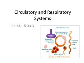
Two circulatory circuits have evolved in vertebrates: • Pulmonary circuit: blood is pumped from heart to lungs and back again • Systemic circuit: blood travels from heart to rest of the body and back to heart Closed vascular system contains: • Arteries carry blood away from heart and branch into arterioles that feed capillary beds • Capillaries aresite of exchange between blood and tissue fluid • Venules drain capillary beds and form veins, which deliver blood back to heart
Fish Fish hearts have two chambers: • One atrium – receives blood from body • One ventricle – receives pumped blood from atrium and sends it to gills Pay attention to # chambers and general blood flow in vertebrates
Crocodilian Heart – 4 chambers, 2 connected aortas Right Atrium (deoxygenated) Ventricle (partially separated) Lungs Right aorta Left Atrium (oxygenated) Ventricle (partially separated) Left Aorta Arterioles and capillaries (becomes deoxygenated)
Lungfish – making transition to land Lungfish have three-chambered hearts adapted to breathe in air as well as water • Lung formed from gut outpouching functions in air • Divided atriumseparates blood into pulmonary and systemic circuits 3 chambers • Bloodstreams stay separate through single ventricle http://www.sheddaquarium.org/images/articles/Australian_Lungfish_Five.JPG
Reptiles (except crocs) • Behavior of reptiles - Activity comes in bursts followed by periods of inactivity • When they aren’t breathing, it would be a waste of energy to send blood to lungs
Reptiles - Crocs Crocodilians have true 4-chambered heart (completely separated ventricle), 2 connected aortas This allows greater control of shuttling blood away from pulmonary circuit when not breathing (submerged)
Birds and Mammals Birds and mammals have four-chambered hearts and separate pulmonary and systemic circuits with the following advantages: • Systemic circuit always receives blood with higher O2 content • Gas exchange is maximized • Circuits can operate at different pressures
Valves prevent backflow of blood: • Atrioventricular valves • lie between atria and ventricles • prevent backflow when ventricles contract • Pulmonary valve and aortic valve (semilunar valves) • lie between ventricles and major arteries • prevent backflow when ventricles relax
Heart Function Cardiac cycle • Both sides of heart contract at same time • first the two atria contract, then the two ventricles • Two phases: • Systole – when ventricles contract • Diastole – when ventricles relax www.monroecc.edu/depts/pstc/backup
Cardiac cycle • 1. atria contract filling ventricles • 2. Ventricle contract shutting AV valves • “lub” • Systole • 3. blood pumped out of ventricles into pulmonary artery and aorta • 4. ventricles relax, aortic and pulmonary valves shut • “dup” • Diastole • 5. ventricles fill with blood as they relax, # 1 is happening
Pacemaker Cells • Resting membrane potential is less negative than other cardiac cells • Less stable • Action potentials are different from other cells • Slower to rise • Broader • Slower to return to resting potential
How do pacemaker cells differ from rest of muscle? • When Na+ and Ca2+ channels open, positive charges flow into cell causing membrane potential to be less negative • Causes action potential (cell “fires”) – membrane less negative • Na+ channels in pacemaker cells are more open, resting potential is LESS negative • Pacemaker cells can fire more readily • When K+ channels open, positive charges flow out and membrane becomes more negative • Cell membrane returns to more negative resting potential when Na+, Ca2+, and K+ balance • Pacemaker cells have unstable resting potential because of the specific behavior of these cation channels • This unstability causes SA node to fire stimulating rest of atria
Nervous system controls heart rate by influencing resting potential: • Norepinephrine from sympathetic nerves increases permeability of Na+/K+ and Ca2+ channels • Resting potential rises more quickly and action potentials are closer together • Acetylcholine from parasympathetic nerves increases permeability of K+ and decreases that of Ca2+ channels – opposite effect of norephinephrine • Resting potentials rise more slowly and action potentials are farther apart
Heart muscle contraction is coordinated: • Action potential is generated in the sinoatrial node (those pacemaker cells) • Action potential spreads through gap junctions in atria both atria contract together • But action potential does not spread to ventricles
Instead, action potential in atria stimulates atrioventricular node • Node consists of non-contracting cells that send action potentials to ventricles via bundle of His • Bundle divides into right and left bundle branches that run to tips of ventricles http://www.univie.ac.at/cga/courses/BE513/EKG/condHeart.gif
From apex, Purkinje fibers spread throughout ventricles • Contraction spreads rapidly and evenly throughout ventricles • Delay between atrial contraction and ventricles ensures proper blood flow
Electrocardiogram • aka ECG or EKG • uses electrodes to record events in cardiac cycle • Large action potentials in heart cause electrical current to flow outward to all body parts • Electrodes register voltage difference at different times
Wave patterns EKG are labeled with letters corresponding to events • P-wave: Atria depolarization • Q,R, and S waves: Ventricular depolarization • T-wave: Relaxation and repolarization of ventricles
Blood pressure & flow through large arteries are high • Flow through capillaries is lower • Pressure is reduced in smaller vessels because: • Arterioles are highly branched larger total cross-sectional area • Capillaries contribute an enormous surface area
Atherosclerosis: “hardening of the arteries” • Endothelial lining of arteries is damaged by high blood pressure, smoking, diet, or microorganisms • Plaque forms at sites of damage • Damaged cells attract migration of smooth muscle cells www.med.uottawa.ca/patho/cardio/ www.cardiocheck.co.uk/mediac/400_0/media/
www.pathguy.com/lectures Figure 50.15 Atherosclerotic Plaque Thrombus, or blood clot, removed from artery