Download
1 / 60
610 likes | 825 Views
Vertebrate Closed Circulatory Systems. Closed circulatory systems Cardiac anatomy & its O 2 supply The myogenic heart & the cardiac cycle Blood pressure Anatomical variations Other ‘hearts’. Hearts. Cardiac cycle – pumping action of the heart Two phases Systole – contraction

E N D
Vertebrate Closed Circulatory Systems • Closed circulatory systems • Cardiac anatomy & its O2 supply • The myogenic heart & the cardiac cycle • Blood pressure • Anatomical variations • Other ‘hearts’
Hearts Cardiac cycle – pumping action of the heart Two phases • Systole – contraction • Blood is forced out into the circulation • Diastole – relaxation • Blood enters the heart
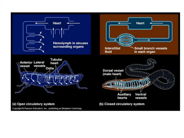
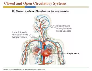
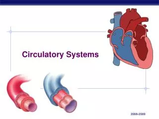
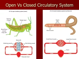
Closed vertebrate circulatory system • Multi-chambered heart • Capillaries connect arterial & venous systems • Respiratory pigments present in red blood cells Tunica media = vascular smooth muscle + elastin fibres Lower BP,thinner walled
Anatomy of the chambered heart bulbus/conus arteriosus Venousbloodpressure • All vertebrates • Similar developmental pathway • Myogenic contractions • Similar intrinsic properties Arterialbloodpressure • Fish: The simplest/earliest design • Four cardiac chambers • All contain muscle (cardiac & smooth) • Surrounded by a pericardial sac • Atrium & ventricle propel blood • Venous BP atrial contraction ventricular contraction • Variations • Hagfishes: incomplete pericardial sac • Sharks & Rays: pericardial sac is stiff; conus arteriosus has cardiac muscle • Primitive Fishes: conus is reduced & bulbus also present • Teleosts: bulbus arteriosus (VSM & elastin fibres)
Closed vertebrate circulatory system • Advantages • Blood pressure can be regulated, even venous blood pressure • High blood pressure, high flow rate & faster circulation time • Exquisite control of blood flow distribution at arterioles (VSM) • High capillary density reduces blood velocity & the diffusion distance to cells • Disadvantages • High resistance to flow b/c of small diameter arterioles (R = r4) • High resistance high blood pressure thicker-walled hearts & higher cardiac O2 needs
Myocardial cells • Striated cells • Electrically connected (desmosomes) • ‘Unstable’ membrane potential Adult mammalian cardiomyocyte Adult fish cardiomyocyte Fish cardiac myocytes also have a reduced sarcoplasmic reticulum (SR), & lack an extensive t-tubular system Consequence: Ca2+ handling during excitation-contraction varies
Myocardium Two types • Compact– tightly packed cells arranged in a regular pattern • Spongy – meshwork of loosely connected cells Relative proportions vary among species • Mammals: mostly compact • Fish and amphibians: mostly spongy • Arranged into trabeculae that extend into the heart chambers
Cardiac muscle O2 supply • A working muscle requires ATP • ATP requirement proportional to cardiac power output • Spongy • Venous blood supply • Simplest, but intricate design • Last organ supplied with O2 • Compact • Coronary blood supply • Compact design • First organ supplied with O2 • Phylogeny & Ontogeny • Hagfishes & Lampreys: spongy • Sharks & Rays: spongy plus variable compact (athletic ability) • Teleosts: most spongy; some have variable compact (athletic/hypoxia) • Amphibians & reptiles: spongy; some have compact (athletic/hypoxia) • Neonatal birds & mammals: spongy • Adult birds & mammals: 99% compact
Cardiac muscle blood & O2 supply Variable compact/spongy Most fish = Trabeculae = venous Mammals = compact = coronary Octopus coronaries
Initiation of cardiac contraction Neurogenic pacemakers: rhythm generated in neurons (some invertebrates) Myogenic pacemakers: rhythm generated in myocytes (vertebrates and some invertebrates) Artificial pacemakers: rhythm generated by device
Control of Contraction • Vertebrate hearts are myogenic – cardiomyocytes produce spontaneous rhythmic depolarizations • Cardiomyocytes are electrically coupled via gap junctions to insure coordinated contractions • Pacemaker – cells with the fastest intrinsic rhythm • Fish: located in the sinus venosus • Other vertebrates: sinoatrial (SA) node in the right atrium
Myogenic contractions • All cardiomyocytes can contract without an external stimulus • Resting membrane potential is ‘unstable’ = Pacemaker potential • Specialised cells (pacemaker) set intrinsic heart rate • Relative timing & speeds of opening of specific ion channels • Increasing heart rate • Norepinephrine is released from sympathetic neurons and epinephrine is released from the adrenal medulla • More Na+ and Ca2+ channels open • Rate of depolarization and action potentials increase • Decreasing heart rate • Acetylcholine is released from parasympathetic neurons • More K+ channels open • Pacemaker cells hyperpolarize • Time for depolarization takes longer
1. Directly between cardiomyocytes • Cardiomyocytes are electrically connected via gap junctions • Electrical signals can pass directly from cell to cell
2. Specialized conducting pathways • Modified cardiomyocytes that lack contractile proteins • Specialized for electrical impulse conduction
Syncitial & sequential cardiac contractions • All cardiomyocytes of a chamber contract together • Electrically coupled cells (desmosomes) • Specialized conduction fibres • Cardiac chambers contract sequentially, after blood has moved • Delays in electrical conduction between chambers (EKG) • Sums all the electrical activity of syncytial contractions & relaxations • P wave: atrial depolarization • QRS complex: ventricular depolarization • T wave: ventricular repolarization
Myogenic contractions • All cardiomyocytes can contract without an external stimulus • But • Different myocardial cells activate different ion channels • Plateau phase – extended depolarization that corresponds to the • refractory period and last as long as the muscle contraction • Prevents tetanus Absence of funny channels Fast Na+ channel Slow L-type Ca2+ channel
Cardiac pumping cycle • ATP muscle contraction blood pressure blood flow • Isometric contraction blood pressure (wall tension) until valves open • Isotonic contraction blood flow (cardiac output) after valves open • Muscle thickness determines pressure
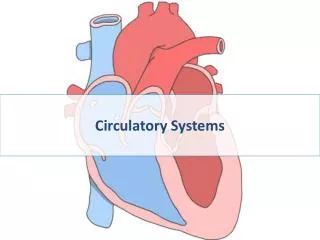
Vertebrate Hearts • Vertebrate hearts have 3 main layers • Pericardium • Myocardium • Endocardium Myocardium
Vertebrate Hearts Have complex walls with four main parts • Pericardium – sac of connective that surround the heart • Two layers: parietal (outer) and visceral (inner) pericardium • Filled with a lubricating fluid • Epicardium – outer layer of heart made of connective tissue • Continuous with visceral pericardium • Contain nerves that regulate the heart • Contain coronary arteries • Myocardium – the middle layer of heart muscle • Endocardium – innermost layer of connective tissue covered by epithelial cells (called endothelium)
Vertebrate hearts - Myocardium • Muscle layer • Composed of cardiomyocytes • Specialized type of muscle cell
Oxygen supply to heart • Myocardium extremely oxidative; has high O2 demand • Coronary arteries supply oxygen to compact myocardium • Spongy myocardium obtains oxygen from blood flowing through the heart
Mammalian cardiac anatomy Two atria Two ventricles
Mammalian cardiac cycle • Step 1: Late diastole, chambers relaxed, passive filling • Step 2: Atrial systole, EDV • Step 3: Isovolumic ventricular contraction • Step 4: Ventricular Ejection • Step 5: Early diastole, semilunar valves close
Electrical and Mechanical Events in the Cardiac Cycle • Heart sounds: opening and closing of valves Figure 9.26
Heart Pressures • The two ventricles contract simultaneously, but the left ventricle contracts more forcefully and develops higher pressure • Resistance in the pulmonary circuit is low due to high capillary density in parallel • Less pressure is needed to pump blood through this circuit • The low pressure also protects the delicate blood vessels of the lungs
Cardiac Output • Cardiac output (CO) – amount of blood the heart pumps per unit time • Stroke volume (SV) – amount of blood the heart pumps with each beat • Heart rate (HR): rate of contraction • CO = HR X SV • Bradycardia – decrease in HR • Tachycardia – increase in HR
Modulating cardiac output • By changing heart rate • By changing stroke volume Concept check: How would you modulate heart rate? Slow heart rate = bradycardia Fast heart rate = tachycardia Stroke volume is regulated in two ways: • Extrinsically (by nervous system and hormones) • Intrinsically (via local mechanisms)
Control of cardiac output: Intrinsic control mechanisms • The importance of cardiac output (Q) • Heart ratePacemaker rate: temperature; body size • Cardiac stroke volume Species variability Effects of filling (venous) pressure
The importance of cardiac output (Q) Flow (output) of blood per unit time from the heart (ml/min/kg) Cardiac power output (= ATP need = O2 need) Power output = Q x [blood pressure developed] Right vs left Atrium vs ventricle
The importance of cardiac output (Q) Respiratory function: O2 uptake = Q x (A-V O2 difference) (Cao2-Cvo2); tissue O2 extraction Q10 effect: O2 uptake doubles for +10oC Species variability in routine & maximum Q values Humans @ 37oC 70-300 ml/min/kg Hagfish @ 10oC 10-30 ml/min/kg Trout @ 10oC 15-50 ml/min/kg Tuna @ 28oC 100-200 ml/min/kg Icefish @ 0oC 100 ml/min/kg Q10 effect~ x8 ~ x8 ~ x2~ x16 [Hb] is a primary determinant of Cao2
Contribution of Q during exercise Human exercising O2 uptake = Q x (A-V O2 difference) Q = [heart rate] x [cardiac stroke volume] Q = 3-fold increaseHR = 2.5-fold increaseSVH = 20% increase A-VO2 = 3-fold increase Volume = O2 delivery to tissues 10-fold increase
Regulation of Q during exercise • Intrinsic pacemaker rate Temperature Body mass • Extrinsic modulation of pacemaker • CNS • Hormones Ions • Intrinsic contractile properties Cardiac stretch Temperature • Extrinsic modulation of contractility • CNS Hormones Ions Q = [heart rate] x [cardiac stroke volume]
Acute temperature effect on heart rate 60 HR,bpm 20 0 20 40 Temperature, oC Ectotherms & Endotherms human trout Cooling by D10oC 2x decrease Q10 ~ 2
Temperature acclimation (resetting of pacemaker rate) 60 HR,bpm 20 0 20 40 Temperature, oC Ectotherms 1. Compensation eg, trout, Q10 = 1-2 trout 2. Downregulation eg, turtles, Q10 > 3 Acute Q10 ~ 2