Download
1 / 50
500 likes | 600 Views
Lecture II. The Nervous System and Its Cells. Bio 3411 Friday August 28, 2009. T. Woolsey 3802 North Building 362-3601 woolseyt@medicine.wustl.edu. Readings. NEUROSCIENCE: 3 rd ed, pp 1-22 THE BRAIN ATLAS: 3 rd ed, pp 4-17 † References:

E N D
Lecture II. The Nervous System and Its Cells Bio 3411 Friday August 28, 2009
T. Woolsey • 3802 North Building • 362-3601 • woolseyt@medicine.wustl.edu Lecture II. The Nervous System and Its Cells
Readings NEUROSCIENCE: 3rd ed, pp 1-22 THE BRAIN ATLAS: 3rd ed, pp 4-17† References: Jellison et al (2004). Diffusion tensor imaging of cerebral white matter: a pictorial review of physics, fiber tract anatomy, and tumor imaging patterns. AJNR Am J Neuroradiol, 25:356-369† Ludwig, E., & Klingler, J. (1956). Atlas cerebri humani. Der innere Bau des Gehirns dargestellt auf Grund makroskopischer Praparate. The inner structure of the brain demonstrated on the basis of macroscopical preparations. Boston,: Little, Brown. Ramón y Cajal, S. (1988). Recollections of my life. New York: Garland. ______________________ †(pdfs on course website: [http://artsci.wustl.edu/~sdanker/index.html]) Lecture II. The Nervous System and Its Cells
Movie - vmjr-brain.mov Lecture II. The Nervous System and Its Cells
Overview • A Few Facts • Main Features of Nervous System • Cells of Nervous System • Importance in Health and in Disease Lecture II. The Nervous System and Its Cells
Facts Lecture II. The Nervous System and Its Cells
OrganWeight 2-3% of bodyO2 Consumption20% of totalBrain Energy (Glucose) Utilization20% of totalBrain Blood Flow20% of heart output at rest Lecture II. The Nervous System and Its Cells
ElementsNeurons (=nerve cells) ≈ 100 BillionGlia (= glue; “supporting” cells) ≈ 1 TrillionSynapses (=clasp) 1/1,000,000th of all stars & planets in the universe/person [less than the total of human synapses of people living in the St.L area!!]Genes 50% of ≈ 20,000-25,000 genes in the human genome are expressed only in Brain[70% of the balance are also expressed in the nervous system; the total is 85% of the genome] Lecture II. The Nervous System and Its Cells
FeaturesBrain, Spinal Cord, Other Lecture II. The Nervous System and Its Cells
THE BRAIN ATLAS 3rded, p. 8 Mid-line (sagittal) section through central nervous system (CNS). Note the relationship between vertebrae (BLACK), segments of the spinal cord (RED) and spinal nerves (YELLOW). Lecture II. The Nervous System and Its Cells
THE BRAIN ATLAS 3rd ed, p. 111 Magnetic Resonance Image (MRI) of head and neck at the midline. Lecture II. The Nervous System and Its Cells
Peripheral (PNS - outside the skeleton)Sensory (sensation) Motor (movement) Autonomic (“involuntary”) Enteric (gut)Central (CNS -inside the skeleton)Spinal Cord (Spine) Brain (Skull) Lecture II. The Nervous System and Its Cells
Views of the human spinal cord and lower brain stem. LEFT - Left lateral (side) showing segments and spinal nerves. MIDDLE - Anterior (front) view of spinal cord without showing enlargements. RIGHT - Posterior (back) view of spinal cord with roots, ganglia and nerves. Lecture II. The Nervous System and Its Cells
THE BRAIN ATLAS 3rd ed, p. 49 Spinal Cord Segment Lecture II. The Nervous System and Its Cells
THE BRAIN ATLAS 3rd ed, p. 20 Left Lateral (side) view of the human Brain Lecture II. The Nervous System and Its Cells
THE BRAIN ATLAS 3rd ed, p. 9 The different regions of the brain from the lateral (side) and median section (middle) human brain. These brain regions are discernable in in all vertebrates and in early embryos. (cerebral cortex = gold; thalamus = blue/purple; midbrain = orange; pons = purple, cerebellum = blue; medulla = red/orange; spinal cord = green) Lecture II. The Nervous System and Its Cells
THE BRAIN ATLAS 3rd ed, p. 58 Lecture II. The Nervous System and Its Cells
THE BRAIN ATLAS 3rd ed, p. 59 Lecture II. The Nervous System and Its Cells
THE BRAIN ATLAS 3rd ed, p. 6 Lecture II. The Nervous System and Its Cells
Components Gray Matter • Cortex, Nuclei or Ganglia (groups of nerve cell bodies and neuropil) generally of similar function • Neuropil - neuronal processes, synapses and glia Lecture II. The Nervous System and Its Cells
Components White Matter • Bundles (groups of myelinated axons [see below] that course in the same direction) • Tracts (also groups of axons (myelinated and un-myelinated but indicates origin, destination and therefore function) Lecture II. The Nervous System and Its Cells
Ludwig, E., & Klingler, J. (1956) Jellison et al (2004) Lecture II. The Nervous System and Its Cells
Cerebrospinal Fluid (CSF) The brain and spinal cord are bathed in a colorless fluid called cerebrospinal fluid (CSF). The fluid is made in chambers in the brain called ventricles (blue). It circulates between all the cells and their processes and in the space between a membrane on the brain surface (called the pia mater) and a membrane that is next to the skull or spine (arachnoid mater) called the subarachnoid space (gold). Lecture II. The Nervous System and Its Cells
Components • Other • Blood Vessels (arteries, capillaries, veins and venous sinuses) • Coverings - meninges (dura mater (tough mother), arachnoid (spider web like), pia (tender/affectionate)) • Cerebrospinal fluid (CSF - ventricles, canals, intercellular space, subarachnoid space) Lecture II. The Nervous System and Its Cells
Movie - vmjr-brain.mov Lecture II. The Nervous System and Its Cells
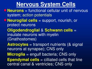
Cells of Nervous SystemNeurons, Contacts, Support Lecture II. The Nervous System and Its Cells
Santiago Ramón y Cajal (1851-1932) ca. 1892 Cajal (say kaahaal) shared the 1906 Nobel Prize for discoveries indicating that the nervous system was made up of individual contiguous elements - the neurons. Lecture II. The Nervous System and Its Cells
Cells of Nervous System Neurons • Parts: cell body (soma), dendrites (input processes), axon (output process) • Types: local circuit (90%), projection (10%) • Variations: stellate (star like); pyramidal (conical/triangular); famous guys - Purkinje, Betz, Cajal, Retzius, Mauthner… • All variations are correlated to particular functions. Lecture II. The Nervous System and Its Cells
Photograph of neurons stained by Golgi’s method which fills processes of some cells with black precipitates of heavy metals and Nissl which stains all nuclei and neuronal cytoplasm blue. Lecture II. The Nervous System and Its Cells
Pyramidal neuron (conical cell body) stained by Golgi’s method. There are multiple processes that resemble branches of trees (dendrites) and one that resembles a wire (axon; arrow). Inputs to the cell are mainly on dendrites and the cell body (soma) while outputs are mainly via the axon. This the principal long axon (output) cell in the cerebral cortex. Lecture II. The Nervous System and Its Cells
Photograph of a Purkinje cell in the cerebellum stained by Golgi’s method. The neuron has one complex dendrite that resembles a sea fan (arrow). Synapses on this cell type are estimated to be about 0.5 Million. Lecture II. The Nervous System and Its Cells
Cells of Nervous System Contacts (Synapses) • Parts:bouton or ending (contains vesicles (transmitters, modulators) and mitochondia), presynaptic membrane (dense in electron microscope); synaptic cleft; postynaptic membrane (dense in electron microscope) • Types:asymmetrical = Type I (postsynaptic membrane is thicker than presynaptic membrane; spherical clear vesicles) these are excitatory synapses - on; symmetrical = Type II (postsynaptic membrane same as presynaptic membrane; flattened clear vesicles) these are inhibitory synapses - off • Variations:large like to muscle, “chalice” in brain stem, “climbing” in the cerebellum; intermediate; small; in passing or as a terminal. All variations relate to specific functions (like variations in hammers - sledge vs. jeweler’s). Lecture II. The Nervous System and Its Cells
Photograph of the giant neuron in the brainstem of the gold fish (Mauthner) stained by Bodian’s method. Synapses on this cell type are particularly easy to see. Much work on this cell type contributed to understanding the structure of the synapse before the electron microscope was perfected. Lecture II. The Nervous System and Its Cells
Electron micrograph of a synapse in the brain stained with the heavy metal element osmium (Os) which is lipophylic (stains lipids/fats). This synapse is only about 2 micrometers across. The main components of a synapse: synaptic cleft (space between the terminal and target process), membrane thickenings on the terminal (pre) and process (post), mitochondria and synaptic vseicles (contain transmitter(s)). Lecture II. The Nervous System and Its Cells
Most brain synapses (type I) have a wider cleft (space between the terminal and target process), thicker membrane densities on the terminal (pre) and process (post) and rounder vesicles. Such synapses are excitatory (on). About 10 - 20% brain synapses (type II) have a narrower cleft, thinner membrane densities on the terminal (pre) and process (post) and flat vesicles. Such synapses are inhibitory (off). Lecture II. The Nervous System and Its Cells
A neuron (red) grown in tissue-culture. Green shows proteins in processes from pre-synaptic neurons. The green/yellow dots on the red neuron indicate synapses. The inset shows a “cartoon” of blue synaptic terminals contacting a neuron. The picture gives a sense of the enormous numbers, distribution and density of synapses on a nerve cell. Neurons “integrate” information from thousands of synapses from many different sources. Lecture II. The Nervous System and Its Cells
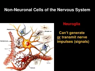
Cells of Nervous System Glia (glue) or Supporting Cells • Parts: cell body (soma) and "short" processes • Types: astrocytes (star like); oligodendorcytes (fewer (oligo) branches (dendrites); microglia (small ones) • Variations: fleshy, fibrous (stringy), myelinating, non-myelinating • All variations relate to specific functions. Lecture II. The Nervous System and Its Cells
Cajal’s drawing of “glia” in the spinal cord. B C A Ependyma (lining of the central canal of the spinal cord) A B Oligodendrocytes which myelinate axons in fiber tracts D C Astrocytes - Protoplasmic (fleshy) in gray matter C D Astrocytes - Fibrous Lecture II. The Nervous System and Its Cells
Astrocyte Oligodendrocyte Microglial Cell NEUROSCIENCE (3rd ed, p.8, fig 1.5) Lecture II. The Nervous System and Its Cells
THE BRAIN ATLAS 3rd ed, pp. 5, 7 Lecture II. The Nervous System and Its Cells
How Does This Work? Please vote on the following proposition:I think that the TA can run up the stairs to the back row of this auditorium in less than 10 seconds. Yes ____ No ____ (Estimated elapsed time: ) Lecture II. The Nervous System and Its Cells
ImportanceBiology, Disease Lecture II. The Nervous System and Its Cells
Biology • Understanding the brain is THE major question in biology and science. • Is it possible for the brain to understand itself? • The brain like any organ has functions: input, output, “thought”, communication. Lecture II. The Nervous System and Its Cells
Brain Diseases • Interfere with brain functions as heart disease interferes with the circulation. • Many diseases have a strong genetic component. • Prevalence is high: ≈ 15 - 30% of the population. • Cost is high: >> $2+ Trillion/year in care, lost income, social services, etc., in the US. • Impact (personal, family, societal) is persistent, pervasive, enormous, incalculable. Lecture II. The Nervous System and Its Cells
What this lecture was about • A Few Facts (genes, size, energy) • Main Features of Nervous System (brain, spinal cord, periphery) • Cells of Nervous System (neurons, glia, contacts) • Importance in Health and in Disease (bases, prevalence, impact) Lecture II. The Nervous System and Its Cells
Movie - vmjr-brain.mov Lecture II. The Nervous System and Its Cells