Download
1 / 62
650 likes | 929 Views
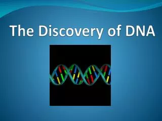
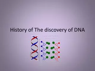
The discovery of DNA. http://www.dnai.org/timeline/index.html. 1860’s: A time of big ideas! Darwin, Mendel (many still debating the cell theory!) Miescher was given the task of researching the composition of lymphoid cells -- white blood cells. (Really gross!)

E N D
The discovery of DNA http://www.dnai.org/timeline/index.html • 1860’s: A time of big ideas! Darwin, Mendel (many still debating the cell theory!) • Miescher was given the task of researching the composition of lymphoid cells -- white blood cells. (Really gross!) • Isolated a white, acidic substance he called nuclein because it was found in the nucleus.
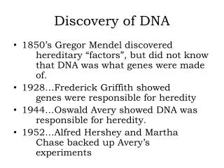
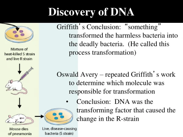
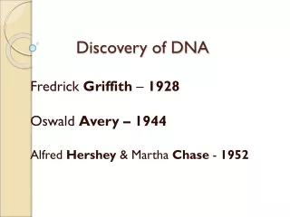
Is DNA the genetic material ? • In addition to circumstantial evidence, two key experiments demonstrated that DNA is the genetic material. • In the first key experiment (Griffiths, 1928) showed that a virulent strain of Streptococcus pneumoniae. genetically transformed nonvirulent S. pneumoniae. into virulent bacteria.
There are two strains of Streptococcus pneumoniae. ROUGH COLONY (R) SMOOTH COLONY (S) S strain is virulent (Polysaccharide capsule prevents detection by host’s immune system) R strain is benign (Lacking a protective capsule, it is recognized and destroyed by host’s immune system)
DNA is the genetic material! • In a prelude to the second key experiment (Avery, 1944) showed that DNA was the transforming agent through studies of T2 bacteriophage and their treatment with hydrolytic enzymes. • The second key experiment (Hershey & Chase, 1952) showed that labeled viruses were incubated with host bacteria. Labeled viral DNA entered host cells, producing many label-bearing viruses. • http://www.dnai.org/timeline/index.html
Many scientists thought that protein was more likely than DNA to be the source of heredity because there are 20 monomers of protein and only 4 types of monomers of DNA. • 1952 - Hershey and Chase did an experiment to determine which one it was
DNA insertion Notice the DNA being inserted into the bacterium
Phage head Sheath Tail fiber DNA 100 nm Bacterial cell
Phage Reproductive Cycle Lytic Cycle (more on this later!)
Chargaff’s Rules • The amounts of A, T, G, and C in DNA: • Identical in identical twins • Varies between individuals of a species • Varies more from species to species • In each species, there are equal amounts of: • A & T • G & C • All this suggests DNA uses complementary base pairing to store genetic info • Human chromosome estimated to contain, on average, 140 million base pairs • Number of possible nucleotide sequences 4,140,000,000
4 Bases = adenine, thymine, guanine, cytosine Pyrimidines - single ring bases Purines - double ring bases
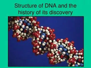
How can we determine the structure of DNA? • X-ray crystallography showed that the DNA molecule is a helix.
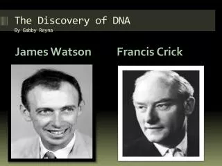
So, what does DNA look like? • Watson and Crick (1953) proposed that DNA is a double-stranded helix with antiparallel strands, and with bases linked by hydrogen bonding. • Their model accounts for genetic information, mutation, and replication functions of DNA. • “A genetic material must carry out two jobs: duplicate itself and control the development of the rest of the cell in a specific way.” • -Francis Crick
Monomer of DNA = Nucleotide Nucleotide = sugar, base, phosphate Backbone of DNA = sugar and phosphates, held together with covalent bonds
The Dawn of Molecular Biology April 25, 1953 Watson and Crick: "It has not escaped our notice that the specific (base) pairing we have postulated immediately suggests a possible copying mechanism for the genetic material.” http://www.cem.msu.edu/~reusch/VirtualText/nucacids.htm
DNA Replication (During S of Interphase!)
DNA Replication • Semiconservative, conservative, and dispersive models for DNA replication were hypothesized. • Each obeyed base-pairing rules. • So, which is it?
DNA Replicationmore history… • Kornberg (1956) demonstrated in vitro that DNA served as its own template during replication. • Meselson and Stahl’s experiment (1957) proved replication of DNA to be semiconservative. A parent strand is a template for synthesis of a new strand. Two replicated DNA helices contain one parent strand and one synthesized strand each.
The Mechanism of DNA Replication • Preview! http://www.wiley.com/college/pratt/0471393878/student/animations/dna_replication/index.html
Complementary • Base pairing • Double Helix is • Essential to DNA’s • Function
The Mechanism of DNA Replication • News Flash: The DNA replication complex is in a fixed location and DNA is threaded through it for replication. • Old idea was via moving replication forks.
DNA Replication con’t. • Prokaryotes have a single origin of replication; eukaryotes have many (102 to 103). Think about the size/amount of DNA in an eukaryotic organism. It would take a long time to replicate the entire genome if it only occurred in one spot! • Bases are added at a rate of 50 per second in a mammal and 500 per second in a bacteria! • Replication for each proceeds in both directions from an origin of replication.
Major protein helpers: • Many proteins assist in DNA replication. • DNA helicases unwind the double helix • Template strands are stabilized by single-stranded binding (SSB’s) proteins. • An RNA primase catalyzes the synthesis of short RNA primers, and to which nucleotides are added. • DNA polymerase catalyzes nucleotides from the 5’ to the 3’ end.
The Mechanism of DNA Replication • DNA polymerase III action causes the leading strand to grow in the 5’-to-3’ direction until replication of that section of DNA is complete. • RNA primer is degraded and DNA is replaced by DNA polymerase I.
The Mechanism of DNA Replication • On the lagging strand, growing in the other direction, DNA is made in the 5’-to-3’ direction but synthesis is discontinuous: DNA is added as short Okazaki fragments to primers, then DNA polymerase III skips past the 5’ end to make the next fragment. • DNA polymerase I and Ligase are required to make lagging strand “continuous”.
Notice the difference between the continuous strand and the lagging strand.
Many Proteins Collaborate at the Replication Fork DNA pol III SSBPs Direction Helicase DNA pol III Primase
Two Daughter Stands form Different Ways Continuous vs. Discontinuous!
Finishing touches on the discontinuous or lagging strand.
The main proteins of DNA replication and their functions III III I I