Download
1 / 12
290 likes | 2.64k Views
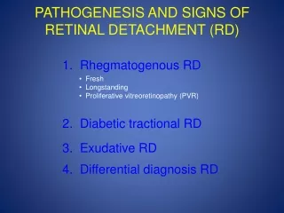
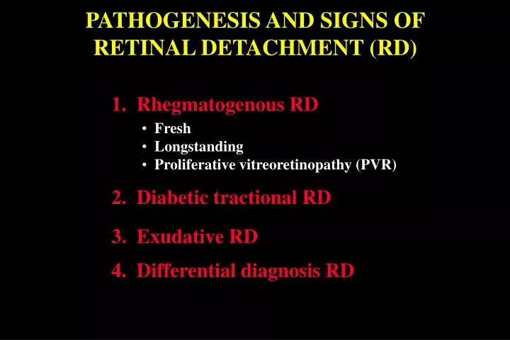
PATHOGENESIS AND SIGNS OF RETINAL DETACHMENT (RD). 1. Rhegmatogenous RD. Fresh Longstanding Proliferative vitreoretinopathy (PVR). 2. Diabetic tractional RD. 3. Exudative RD. 4. Differential diagnosis RD. Pathogenesis of rhegmatogenous RD.

E N D
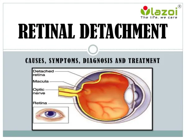
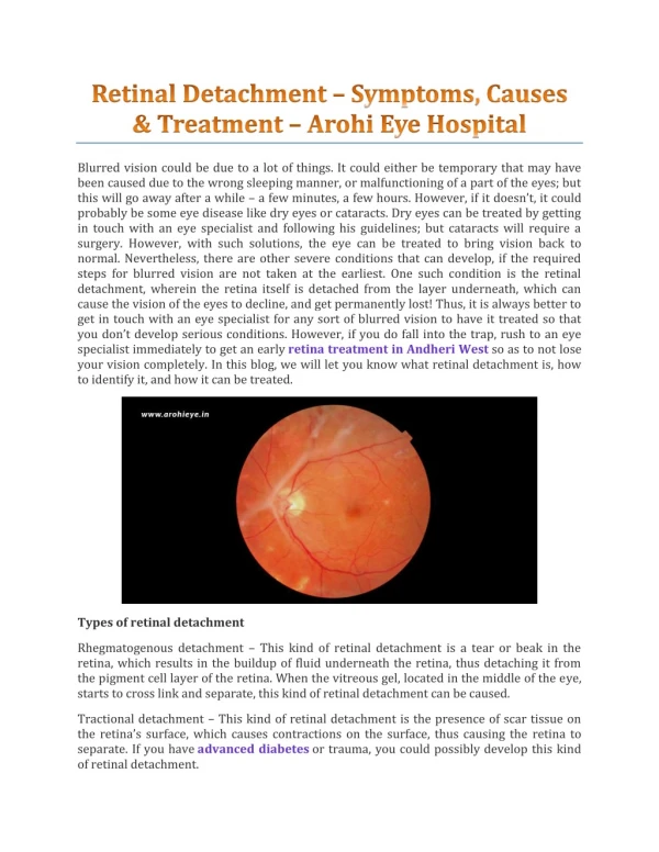
PATHOGENESIS AND SIGNS OF RETINAL DETACHMENT (RD) 1. Rhegmatogenous RD • Fresh • Longstanding • Proliferative vitreoretinopathy (PVR) 2. Diabetic tractional RD 3. Exudative RD 4. Differential diagnosis RD
Pathogenesis of rhegmatogenous RD • Two components for retinal break formation • Acute posterior vitreous detachment (PVD) • Predisposing peripheral retinal degeneration Possible sequelae of acute PVD Retinal tear formation and haemorrhage (10-15%) Avulsion of retinal vessel and haemorrhage (uncommon) Uncomplicated PVD (85%)
Fresh rhegmatogenous RD - signs • Annual incidence - 1:10,000 of population • Eventually bilateral in 10% • Loss of choroidal pattern • Retinal breaks • Convex, deep mobile elevation • extending to ora serrata • Slightly opaque with dark blood vessels
Longstanding rhegmatogenous RD - signs • Frequently inferior with small holes • Very thin retina • Secondary intraretinal cysts • Demarcation lines (high-water marks)
Proliferative vitreoretinopathy Grade A (minimal) Grade B (moderate) Grade C (severe) • Vitreous haze and • tobacco dust • Retinal wrinkling and • stiffness • Rolled edges of tears • Rigid retinal folds • Vitreous condensations • and strands
Pathogenesis of diabetic tractional RD (1) Antero-posterior traction RD Preretinal haemorrhage
Pathogenesis of diabetic tractional RD (2) A-P traction Bridging traction Preretinal haemorrhage
Signs of diabetic tractional RD • Slow progression and variable fibrosis • Does not extend to ora serrata • Concave, shallow immobile elevation • Highest at sites of vitreoretinal traction
Pathogenesis and Causes of Exudative RD • Damage to RPE by subretinal disease • Passage of fluid derived from choroid into subretinal space 1. Choroidal tumours • Primary • Metastatic 2. Intraocular inflammation • Harada disease • Posterior scleritis 3. Systemic • Toxaemia of pregnancy • Hypoproteinaemia 4. Iatrogenic • RD surgery • Excessive retinal photocoagulation 5. Miscellaneous • Choroidal neovascularization • Uveal effusion syndrome
Signs of exudative RD • Convex, smooth elevation • May be very mobile and deep with • shifting fluid • Subretinal pigment (leopard spots) • after flattening
Differential diagnosis of RD Degenerative retinoschisis Choroidal detachment Uveal effusion syndrome • Frequently bilateral • Smooth, thin and immobile • Occasionally breaks in one • or both layers • Associated with hypotony • Unilateral, brown, smooth, • solid and immobile • Ora serrata may be visible • Idiopathic • Rare, unilateral • Combined choroidal and • exudative detachments