Download

1 / 29
330 likes | 798 Views
Learn about the different types of retinal detachment, its pathogenesis, signs, and treatment options including prophylaxis methods. Discover the risk factors and symptoms associated with this condition.

E N D
PATHOGENESIS AND SIGNS OF RETINAL DETACHMENT (RD) 1. Rhegmatogenous RD • Fresh • Longstanding • Proliferative vitreoretinopathy (PVR) 2. Diabetic tractional RD 3. Exudative RD 4. Differential diagnosis RD
Definition and classification • Break - full-thickness defect in sensory retina • Hole - caused by chronic retinal atrophy • Tear - caused by dynamic vitreoretinal traction Morphology of tears a. Complete U-tear b. Linear c. Incomplete L-shaped d. Operculated e. Dialysis
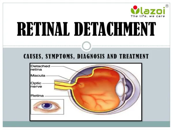
Retinal detachment (RD) Separation of sensory retina from RPE by subretinal fluid (SRF) Rhegmatogenous - caused by a retinal break Non-rhegmatogenous - tractional or exudative
Indirect ophthalmology Condensing lenses Technique • Keep lens parallel to patient’s iris plane • Avoid tendency to move towards patient • Ask the patient to move eyes and head • into optimal positions for examination • The higher the power, the less the • magnification, the shorter the working • distance but the greater the field of view
Fundus drawing Technique Colour code Breaks Detached retina Vitreous opacity Thinning Exudate Lattice Retinal pigment • Place chart upside down • Draw what you see
Pathogenesis of rhegmatogenous RD • Two components for retinal break formation • Acute posterior vitreous detachment (PVD) • Predisposing peripheral retinal degeneration Possible sequelae of acute PVD Retinal tear formation and haemorrhage (10-15%) Avulsion of retinal vessel and haemorrhage (uncommon) Uncomplicated PVD (85%)
Retinal breaks not requiring treatment g - Small asymptomatic holes near ora serrata e - Asymptomatic dialysis surrounded by pigment h - Small inner layer holes in retinoschisis f - Breaks in both layers of retinoschisis
Complications of lattice degeneration • No complications - in most cases • RD associated with atropic holes, particularly in young myopes • RD associated with tractional tears in eyes with acute PVD Indications for prophylaxis • RD in fellow eye • Extensive lattice in high myopia
PROPHYLAXIS OF RHEGMATOGENOUS RETINAL DETACHMENT 1. Retinal breaks 2. Predisposing degenerations • Lattice • Snailtrack • White-without-pressure 3. Treatment modalities • Laser photocoagulation • Cryotherapy 4. Benign peripheral degenerations
pulled away from the underlying choroid • small areas of the retina torn => retinal tears or retinal breaks • retinal cells deprived of oxygen • if not promptly treated =>permanent vision loss
Fresh rhegmatogenous RD - signs • Annual incidence - 1:10,000 of population • Eventually bilateral in 10% • Convex, deep mobile elevation • extending to oraserrata • Slightly opaque with dark blood vessels • Loss of choroidal pattern • Retinal breaks Longstanding rhegmatogenous RD - signs • Frequently inferior with small holes • Very thin retina • Secondary intraretinal cysts • Demarcation lines (high-water marks)
Proliferative vitreoretinopathy Grade A (minimal) Grade B (moderate) Grade C (severe) • Vitreous haze and • tobacco dust • Retinal wrinkling and • stiffness • Rolled edges of tears • Rigid retinal folds • Vitreous condensations • and strands Signs of diabetic tractional RD • Concave, shallow immobile elevation • Highest at sites of vitreoretinal traction • Slow progression and variable fibrosis • Does not extend to ora serrata
Pathogenesis of diabetic tractional RD (1) Antero-posterior traction RD Preretinal haemorrhage
Pathogenesis of diabetic tractional RD (2) A-P traction Bridging traction Preretinal haemorrhage
Pathogenesis and Causes of Exudative RD • Damage to RPE by subretinal disease • Passage of fluid derived from choroid into subretinal space 1. Choroidal tumours • Primary • Metastatic 2. Intraocular inflammation • Harada disease • Posterior scleritis 3. Systemic • Toxaemia of pregnancy • Hypoproteinaemia 4. Iatrogenic • RD surgery • Excessive retinal photocoagulation 5. Miscellaneous • Choroidal neovascularization • Uveal effusion syndrome
Signs of exudative RD • Convex, smooth elevation • May be very mobile and deep with • shifting fluid • Subretinal pigment (leopard spots) • after flattening
SYMPTOMS • floaters • light flashes • shadow or curtain over a portion of visual field • blur in vision
Can occur as a result of: • trauma • advanced diabetes • an inflammatory disorder, such as sarcoidosis • shrinkage of the jelly-like vitreous that fills the inside of the eye
Factors that may increase risk of retinal detachment: • aging - more common in people older than 40 • previous retinal detachment in one eye • family history of retinal detachment • extreme nearsightedness • previous eye surgery • previous severe eye injury or trauma
TREATMENTS Retinal tears: • laser surgery (photocoagulation) • freezing (cryopexy) Retinal detachment: • pneumatic retinopexy • scleral buckling • vitrectomy
Technique of cryotherapy • Surround lesion with single row of • cryo-applications • Preferred for treatment of large • areas
PRINCIPLES OF RETINAL DETACHMENT SURGERY 1. Scleral buckling • Configuration of buckles • Preliminary steps • Localization of breaks • Cryotherapy • Insertion of local explant • Encircling procedure • Drainage of subretinal fluid • Causes of early failure 2. Pneumatic retinopexy 3. Vitrectomy • Giant tears • Proliferative vitreoretinopathy (PVR) • Diabetic tractional RD
Configuration of scleral buckles Segmental circumferential Radial Encircling augmented by radial sponge Encircling augmented by solid silicone tyre
Drainage of subretinal fluid Indications • Difficulty in localizing break • Immobile retina • Longstanding RD • Inferior RD Complications Haemorrhage Retinal incarceration
Indications RD with superior breaks Pneumatic retinopexy Technique (a) Cryotherapy (b) Gas injection (c) Postoperative positioning (d) Flat retina
Vitrectomy for PVR • Dissection of star folds and peeling of membranes • Injection of expanding gas or silicone oil Vitrectomy for diabetic tractional RD Release of circumferential traction Release of antero- posterior traction Endophotocoagulation