Download
1 / 68
680 likes | 696 Views
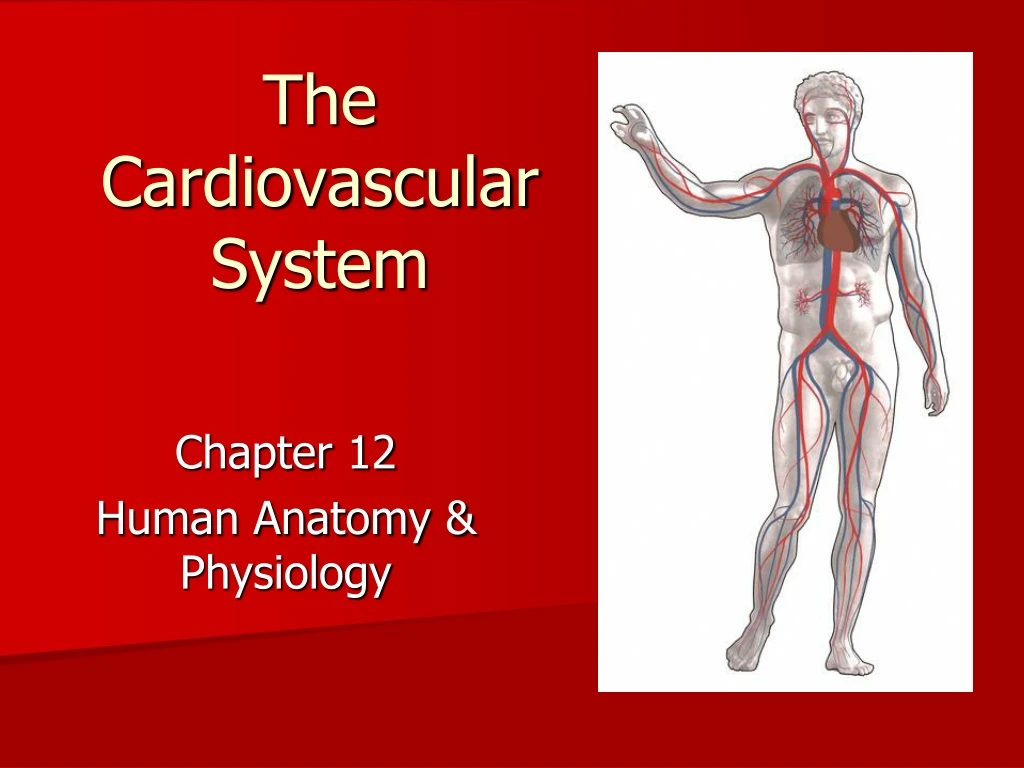
The Cardiovascular System. Chapter 12 Human Anatomy & Physiology. Standard 34.

E N D
The Cardiovascular System Chapter 12 Human Anatomy & Physiology
Standard 34 • Outline the structure and functions of the anatomy of the cardiovascular system, paying special attention to the musculature of the walls, the chambers, and the valves of the heart and blood vessels. Locate and demonstrate the circulation of blood through the heart; describe the phases and importance of the cardiac cycle and how heart rate and cardiac output relate to one another. Listen to heart sounds, either digitally or with a stethoscope, to identify the normal and abnormal sounds made during the cardiac cycle. Explain the causes for abnormal sounds encountered.
Objectives • Describe the anatomy of the heart.
I. Overview of the Cardiovascular System • The circulatory system can be thought of as the transport system of the body. • A closed system consisting of the heart,blood vessels, and blood • The heart pumps blood • Blood vessels allow blood to circulate to all parts of the body • Function: Deliver oxygen & nutrient-rich blood to body cells and remove carbon dioxide and waste
A. Overview of the Heart • The heart is located in the thoracic cavity between the lungs slightly to the left • A hollow, cone-shaped muscle about the size of a fist • Made up of cardiac muscle • https://www.youtube.com/watch?v=X9ZZ6tcxArI • https://www.youtube.com/watch?v=DAXa4eR1s0M
II. Anatomy of the Heart 1. Coverings: • Pericardium – a double serous membrane • Visceral pericardium (epicardium) • Next to heart • Parietal pericardium • Outside layer • Serous fluid fills the space between the layers of pericardium
2. Heart Walls: • Three layers a. Epicardium • Outside layer • This layer is the visceral pericardium b. Myocardium • Middle and thickest layer • Mostly cardiac muscle c. Endocardium • Inner layer • Made up of simple squamous epithelium
3. Chambers • The heart has 4 chambers • Left & right atria – receive blood • Left & right ventricles -pump blood out • Chambers are separated by a septum
4. Heart Valves • Valves are flaps of connective tissue between the atria and ventricles • Moves the blood through the heart in one direction • Valves open as blood is pumped through • Held in place by chordae tendineae (“heart strings”) • Valves are closed to prevent backflow
Four valves a. Atrioventricular valves – between atria and ventricles • left atrium bicuspid valve (mitral valve) left ventricle • right atria tricuspid valve right ventricle b. Semilunar valves - between ventricle and artery • right ventricle pulmonary semilunar valve pulmonary ARTERY • left ventricle aortic valve aorta
5. Major Vessels • Aorta – largest artery • Carries blood from left ventricle towards body • Pulmonary arteries • Carries Oxygen-poor blood from the right ventricle to lungs • Vena cava • Superior and inferior • Receives blood from the upper & lower body • Pulmonary veins (4) • Carries Oxygen-rich blood from lungs to the left atrium
5. Major Vessels Con’t • Coronary arteries - supply the heart muscle with oxygen rich blood • Coronary sinus - collection of veins; drains less oxygenated blood from heart muscle to the right atrium
IV. Anatomy of Blood Vessels • Blood Vessels are tubes that transport blood A. Function: • Transport blood • Carry out the exchange of gases and waste • Regulate blood pressure • Direct blood flow
B. Types of Blood Vessels 1. Arteries • Blood vessels which carry oxygen-rich blood away from the heart to the body. • The aorta is the largest artery in our body • Thick walls
2.Capillaries • Small blood vessels which connect arteries and veins together • Where exchange of oxygen, carbon dioxide, nutrients, and waste occur • One cell layer thick
3. Veins • Blood vessels which carry oxygen-poor blood from the body back to the heart. • Thin walls • Valves
Bell Work • Which side of the heart carries oxygenated blood? Deoxygenated?
Objectives • Trace the flow of the blood through the heart.
The Heartbeat • Heart Surgery • https://www.youtube.com/watch?v=tRFMEeQCkpA • https://www.youtube.com/watch?v=Gnv54V8Jj1U
Circulation of Blood in the Body • Movement of Blood Through Vessels • Most arterial blood is pumped by the heart • Veins use the milking action of muscles to help move the blood
Circulation of Blood in the Body B. The goal is to • Send oxygen-poor blood to the lungs to pick up oxygen and then • To pump oxygen-rich blood from the heart to the body cells
B. Three circulation pathways • Pulmonary circulation • From heart to lungs • Systemic circulation • From heart to body • Coronary circulation • From heart to heart muscle
Blood flow through heart https://www.youtube.com/watch?v=H04d3rJCLCE
Blood Flow Through The Heart Deoxygenated Blood From The Body Superior and Inferior Vena Cava Right Atrium Tricuspid Valve R Ventricle Pulmonic Valve Pulmonary Arteries Lungs (Oxygenation Occurs) Pulmonary Veins Left Atrium bicuspid (mitral) valve left ventricle Aortic Valve Aorta Body
Superior Vena Cava Inferior Vena Cava Pulmonary Arteries <- To Lungs To Lungs -> Pulmonic Valve Right Atrium Tricuspid Valve Right Ventricle
Superior Vena Cava Inferior Vena Cava Aorta Pulmonary Arteries <- To Lungs To Lungs -> Aortic Valve Left Atrium Pulmonary Veins Pulmonary Veins Pulmonic Valve Mitral Valve Right Atrium Tricuspid Valve Left Ventricle Right Ventricle
Objectives • Explain the Cardiac Conduction System.
A Cardiac Cycle • One cycle • atria contract / ventricles relax • ventricles contract / atria relax • Systole - contraction phase • Diastole - relaxation phase
Cardiac Cycle • Stroke volume is the volume of blood ejected from one ventricle with each beat • Cardiac output is amount of blood that one ventricle can pump each minute—average is about 5 L per minute at rest
The Conduction System of the Heart https://www.youtube.com/watch?v=fZT9vlbL2uA
Electrical Impulses Through the Heart • SA (sinoatrial) node, the pacemaker—located in the wall of the right atrium near the opening of the superior vena cava • AV (atrioventricular) node—located in the right atrium along the lower part of the interatrial septum • AV bundle (bundle of His)—located in the septum of the ventricle • Purkinje fibers—located in the walls of the ventricles
Measuring the Cardiac Cycle • Electrocardiograms (EKG or ECG) are used to measure the electrical rhythm of the heart’s contraction
Conduction System of the Heart • The normal ECG has three deflections or waves called the P wave, the QRS complex, and the T wave • P wave—associated with depolarization of the atria • QRS complex—associated with depolarization of the ventricles • T wave—associated with repolarization of the ventricles
Conduction System of the Heart • Cardiac dysrhythmia—abnormality of heart rhythm • Heart block—conduction of impulses is blocked • Complete heart block—impaired AV node conduction, producing complete dissociation of P waves from QRS complexes • Can be treated by implanting an artificial pacemaker
Pacemaker • Used to maintain a consistent heart rate when the body’s natural pacemaker (SA node) is not properly functioning
Conduction System of the Heart • Bradycardia—slow heart rate (under 60 beats/min) • Tachycardia—rapid heart rate (over 100 beats/min) • Sinus dysrhythmia—variation in heart rate Fibrillation—condition in which cardiac muscle fibers are “out of step,” producing no effective pumping action https://www.webmd.com/heart-disease/atrial-fibrillation/video/atrial-fibrillation
Heart Failure • Heart failure—inability to pump enough returned blood to sustain life; it can be caused by many different heart diseases • Right-sided heart failure—failure of the right side of the heart to pump blood, usually because the left side of the heart is not pumping effectively
Heart Failure • Left-sided heart failure (congestive heart failure)—inability of the left ventricle to pump effectively, resulting in congestion of the systemic and pulmonary circulations • Diseased hearts can be replaced by donated living hearts (transplants) or by artificial hearts (implants), although both procedures have yet to be perfected
Bell Work • What does each of the following represent? P Wave QRS Complex T Wave
Objectives • Discuss characteristics and treatment of common cardiac and circulatory abnormalities.
Abnormalities 1. Arteriosclerosis • blockage of the arterial walls due to the build up of cholesterol • Leads to hypertension, heart attack, & stroke