Download
1 / 48
480 likes | 501 Views
Unravel the building blocks of DNA and RNA, dissecting their primary and secondary structures, base pairings, and genetic information flow mechanisms. Learn about the molecular biophysics of DNA, topoisomerases, and restriction endonucleases. Dive into the world of nucleic acids, their biotechnological applications, and the impact of genetic diseases such as cystic fibrosis. Understand the relevance of DNA in drug design, cancer research, and genetic typing. Join us to explore the intricate world of DNA through this comprehensive course!

E N D
Biochemistry of Medicinals I – Nucleic Acids Instructor: Natalia Tretyakova, Ph.D. 760E CCRB (Cancer Center) Tel. 6-3432 e-mail trety001@umn.edu Lecture: MWF 3:35-4:25 7-135 WDH Recitation: Th. 11-12 Web page: see “Web enhanced courses”
Chapter 1. DNA Structure. Required reading: Stryer 5th Edition p. 117-125, 144-146, 152, 746-750, 754-762, 875-877) (or Stryer’s Biochemistry 4th edition p. 75-77,80-88, 119-122, 126-128, 787-799, 975-980)
DNA Structure: Chapter outline • Biological roles of DNA. Flow of genetic information. • Primary and secondary structure of DNA. • Types of DNA double helix. Sequence-specific DNA recognition by proteins. • Biophysical properties of DNA. • DNA topology. Topoisomerases. • Restriction Endonucleases. Molecular Cloning
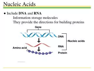
(ribonucleic acids) (deoxyribonucleic acids) replication transcription translation DNA
Why ? • Questions? • How is genetic information transmitted to progeny cells? • How is DNA synthesis initiated? • What causes DNA defects and what are their biological an physiological consequences? • What causes the differences between cells containing the same genetic information? • Relevance: • •Cancer: ex., Xeroderma pigmentosum • •Genetic diseases: ex., cystic fibrosis, sickle cell anemia, inborn errors of metabolism • •Genetic typing: ex., drug metabolism • •Rational drug design: ex., antitumor and antimicrobial drugs • •Biotechnology: ex., growth hormones
The Building Blocks of DNA -N-glycosidic bond
DNA and RNA nucleobases (DNA only) (RNA only)
Example: nucleobase Adenine
Nucleoside 2’-deoxyadenosine
Nucleotide 2’-deoxyadenosine-5’-monophosphate
nucleobase (Deoxy) nucleoside 5’-mononucleotide Adenine (A) Guanine (G) Thymine (T) Cytosine (C) Uracil (U) 2’-Deoxyadenosine (dA) 2’- Deoxyguanosine (dG) 2’- Deoxythymidine (dT) 2’- Deoxycytidine (dC) Uridine (U) Deoxyadenosine 5’-monophosphate (5’-dAMP) Deoxyguanosine 5’-monophosphate (5’-dGMP) Deoxythymidine 5’-monophosphate (5’-dTMP) Deoxycytidine 5’-monophosphate (5’-dCMP) Uridine 5’-monophosphate (5’-UMP) Nomenclature of nucleobases, nucleosides, and mononucleotides
Preferred conformations of nucleobases and sugars in DNA and RNA Sugar puckers: 5.9 A 7.0 A
Nucleosides Must Be Converted to5’-Triphosphates to be Part of DNA and RNA
DNA isArranged5’ to 3’Connected byPhosphates Linking inDNA biopolymer: DNA primary structure
DNA secondary structure – double helix • James Watson and Francis Crick, 1953- proposed a model for DNA structure • DNA is the molecule of heredity (O.Avery, 1944) • X-ray diffraction (R.Franklin and M. Wilkins) • E. Chargaff (1940s) G = C and A = T in DNA Francis Crick Jim Watson
Watson-Crick model of DNA was based on X-ray diffraction picture of DNA fibres (Rosalind Franklin and Maurice Wilkins) Rosalind Franklin
Watson-Crick model of DNA was consistent with Chargaff’s base composition rules Erwin Chargaff (Columbia University) G = C and A = T in DNA
Living Figure – B-DNA http://bcs.whfreeman.com/biochem5
Forces stabilizing DNA double helix • Hydrogen bonding (2-3 kcal/mol per base pair) • Stacking (hydrophobic) interactions • (4-15 kcal/mol per base pair) • 3. Electrostatic forces.
B-DNA • •Sugars are in the 2’ endo conformation. • •Bases are the anti conformation. • •Bases have a helical twist of 36º • (10 bases per helix turn) • Helical pitch = 34 A 23.7 A right handed helix • helical axis passes through • base pairs 7.0 A • planes of bases are nearly • perpendicular to the helix axis. • 3.4 A rise between base pairs Wide and deep Narrow and deep
DNA can deviate from Ideal Watson-Crick structure • Helical twist ranges from 28 to 42° • Propeller twisting 10 to 20° • Base pair roll
N NH 2 H N O 2 N N HN C-1’ N N NH O 2 C-1’ Major and minor groove of the double helix O N NH N N N N C-1’ O C-1’ Wide and deep Narrow and deep
Major groove and Minor groove of DNA N NH O 2 N H N O 2 N NH N N N HN C-1’ N N N N C-1’ NH O O 2 C-1’ Hypothetical situation: the two grooves would have similar size if dR residues were attached at 180° to each other To deoxyribose-C1’ C1’ -To deoxyribose C-1’
B-type duplex is not possible for RNA steric “clash”
A-form helix:dehydrated DNA; RNA-DNA hybrids • •Sugars are in the 3’ endo conformation. • •Bases are the anti conformation. • •11 bases per helix turn • Helical pitch = 25.3 A Right handed helix • planes of bases are tilted • 20 ° relative the helix axis. • 2.3 A rise between base pairs 25.5 A Top View
Living Figure – A-DNA http://bcs.whfreeman.com/biochem5
The sugar puckering in A-DNA is 3’-endo 5.9 A 7.0 A
A-form helix:dehydrated DNA; RNA-DNA hybrids • •Sugars are in the 3’ endo conformation. • •Bases are the anti conformation. • •11 bases per helix turn • Helical pitch = 25.3 A Right handed helix • planes of bases are tilted • 20 ° relative the helix axis. • 2.3 A rise between base pairs 25.5 A Top View
A-DNA has a shallow minor groove and a deep major groove • • Helix axis A-DNA B-DNA
Z-form double helix:polynucleotides of alternating purines and pyrimidines (GCGCGCGC) at high salt • • Backbone zig-zags because sugar puckers alternate between 2’ endo pyrimidines and 3’ endo (purines) • • Bases alternate between anti (pyrimidines) and syn conformation (purines). • •12 bases per helix turn • Helical pitch = 45.6 A Left handed helix • planes of the bases are • tilted 9° relative the helix • axis. • 3.8 A rise between base pairs 18.4 A • Flat major groove • Narrow and deep minor groove
Sugar and base conformations in Z-DNA alternate: 5’-GCGCGCGCGCGCG 3’-CGCGCGCGCGCGC C:sugar is 2’-endo, base is anti G: sugar is 3’-endo, base is syn
Living Figure – Z-DNA http://bcs.whfreeman.com/biochem5
Biological relevance of the minor types of DNA secondary structure • Although the majority of chromosomal DNA is in B-form, • some regions assume A- or Z-like structure • Runs of multiple Gs are A-like • The upstream sequences of some genes contain • 5-methylcytosine = Z-like duplex • Structural variations play a role in DNA-protein interactions • RNA-DNA hybrids and ds RNA have an A-type structure
Hydrogen bond donors and acceptors in DNA grooves facilitate its recognition by proteins H N H N 2 2 The edges of base pairs displayed to DNA major and minor groove contain potential H-bond donors and acceptors: O N n h o h n= Nitrogen hydrogen bond acceptor o= Oxygen hydrogen bond acceptor h= Amino hydrogen bond donor
Hydrogen bond donors and acceptors on each edge of a base pair
Structural characteristics of DNA facilitating DNA-Protein Recogtnition • Major and major groove of DNA contain sequence- • dependent patterns of H-bond donors and acceptors. • Sequence-dependent duplex structure (A, B, Z, bent • DNA). • Hydrophobic interactions via intercalation. • Ionic interactions with phosphates.
Leucine zipper proteins bind DNA major groove Groove binding drugs and proteins 5’-ATT-3’ Others: netropsin, distamycin, Hoechst 33258
Triple helix and Antigene approach Hoogsteen base pairing = parallel Reversed Hoogsteen = antiparallel
Biophysical properties of DNA • Facile denaturation (melting) and re-association of the duplex • are important for DNA’s biological functions. • In the laboratory, melting can be induced by heating. Single strands T° duplex • Hybridization techniques are based on the affinity of complementary • DNA strands for each other. • Duplex stability is affected by DNA length, % GC base pairs, ionic strength, the presence of organic solvents, pH • Negative charge – can be separated by gel electrophoresis
Separation of DNA fragments by gel electrophoresis Polyacrylamide gel: • DNA strands are negatively charged – • migrate towards the anode • Migration time ~ ln (number of base • pairs)