Download
1 / 35
350 likes | 359 Views
Join Jonathan Reynolds, PT, PhD, for a detailed examination, diagnosis, and treatment of common foot and ankle injuries in dancers, including posterior ankle pain and plantar fasciitis. Learn about diagnostic techniques and therapeutic approaches to improve dancer performance and prevent future injuries.

E N D
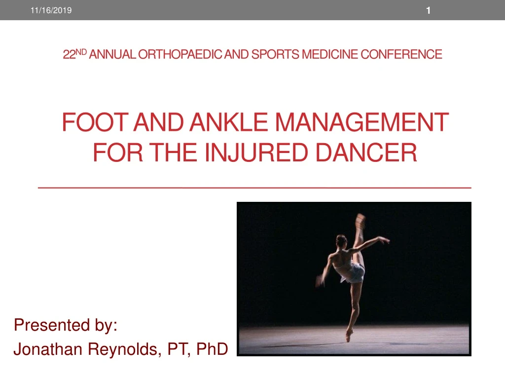
22nd Annual Orthopaedic and Sports Medicine Conference Foot and Ankle Management for the injured dancer Presented by: Jonathan Reynolds, PT, PhD
Diagnostic Excellence • Detailed history • Thorough examination • Radiology dependence • Three major concerns: • Posterior pain • Plantar foot pain • Recruitment/poor co-ordination • Practicum • Examination • Treatment
Posterior Ankle Pain • Achilles tendinitis • Posterior impingement • Talocrural joint • OsTrigonum • Steida’s Process • FHL Tendinitis • Talar stress fracture/OCD • Talocrural malalignment • Anterior translation • Posterior translation
Achilles Tendinitis • Pain at: • Anterior aspect (soleus) of Achilles tendon • Pathomechanics • Overuse (jumping, releve, running) • Evaluation • Provocative tests: • Heel raise • Jump • Soleus stretch • MMT: Up on toes • PROM: ankle DF • Palpation: • Tenderness at soleus aspect of AT • Check teno-periosteal junction at calcaneus Differentiators: Surface anatomy Localization of pain Range of ankle motion when symptoms are felt
Posterior Impingement • Pain at: • Posterior TCJ • Pathomechanics • Overuse (dance, high heels) • OsTrigonum • Steida’s Process • Evaluation • Provocative tests: • Heel raise – pain at EOR • MMT: Up on toes • PROM: ankle PF • Palpation: tenderness at TCJ – posterior capsule Hamilton, W.G., Clin. Sports Med., 1988 • Differentiators: • Pain on forced PF • Patients with PI will OVERUSE gastroc./soleus/AT to compensate → Achilles tendinitis
Posterior Impingement En pointe Releve
Posterior Impingement OsTrigonum
Posterior Impingement Ganglion Cyst Talar Stress Fracture
Flexor Hallucis Longus Tendinitis • Pain at: • Plantar MTPJ • Medial longitudinal arch • Antero-medial calcaneus (?PF) • Retro-malleolar (medial) • Pathomechanics • Forefoot varus → Ankle valgus • Knee valgus • ↓Tissue compliance • Shortened use (ballet, high-heels) • Overuse (jumping, up on toes) • Evaluation • MMT: *1st MT plantarflexion • PROM: *ankle DF with 1st MT DF • Palpation: *tenderness along FHL Differentiators: Surface anatomy MMT of 1st MT Test-treat-re-test
Talar Stress Fracture and OCD • Pain at: • Posterior TCJ • Pathomechanics • Overuse (jumping, running) • Evaluation • Provocative tests: • Forced PF • Downhill walk • MMT: N/A • PROM: ankle PF • Palpation: • Tenderness at: • Posterior TCJ Differentiator: Catching during running and walking
Talocrural Malalignment • Pain at: • Retro-malleolar • Posterior TCJ • Pathomechanics • Repetitive deceleration • Evaluation • Provocative tests: • Forced PF • Pointe • MMT: N/A • PROM: ankle PF • Palpation: N/A • Mixed: talus PA glide is: • Therapeutic • Diagnostic Differentiators: Negative MMT + forced PF
Normal Ankle Mechanics - Dorsiflexion • Talus – glides posteriorly, externally rotates and tilts laterally • Fibula – supero-posterior glide, lateral translation • Proximal TFJ – Fibula moves anterolaterally and superiorly and rotates. • Peroneus longus – plantarflexes 1st ray • Peroneals (longus and brevis) – transfer weight from lateral to medial forefoot. Denegar and Miller, 2002)
Normal Ankle Mechanics - Plantarflexion • Talus – glides anteriorly, internally rotates and tilts medially • Fibula – infero-anterior glide, medial translation • Proximal TFJ – Fibula glides • Superoposteromedially with pronation, and • Inferoanterolaterally with supination. Denegar and Miller, 2002
Plantar Fasciitis • Pain at: • Tenoperiostial junction (TPJ) • Pathomechanics • Forefoot varus →Pronation • Knee valgus • Overload • ↓myofascial compliance • Tissue adaptation • Thickening • Inflammation • Pain • ↑Water-binding capacity • Examination • Alignment: • Hindfoot • Forefoot • Standing posture: • Knee • Foot and ankle • PROM: Tight Achilles/gastroc/soleus • Palpation: greatest at TPJ
Flexor Hallucis Longus Tendinitis • Pain at: • Plantar MTPJ • Medial longitudinal arch • Antero-medial calcaneus (?PF) • Retro-malleolar (medial) • Pathomechanics • Forefoot varus → Ankle valgus • Knee valgus • ↓Tissue compliance • Shortened use (ballet, high-heels) • Overuse (jumping, up on toes) • Evaluation • MMT: *1st MT plantarflexion • PROM: *ankle DF with 1st MT DF • Palpation: *tenderness along FHL Differentiators: Surface anatomy MMT of 1st MT Test-treat-re-test
Sesamoiditis • Pain at: • Plantar aspect of first ray • Pathomechanics • Forefoot varus • Knee valgus • Tight/overactive hip adductors • Weak/inhibited hip abductors and external rotators • Poor myofascial compliance • Shortened use (ballet, Irish dance high-heels) • Overuse (jumping, up on toes) • Evaluation • Provocative test: • Heel raise • Hopping • MMT: 1st MT plantarflexion • PROM: ankle DF with 1st MT DF • Palpation: *tenderness at sesamoids Differentiators: Surface anatomy Localized pain
Chronic Ankle Instability (CAI) • Alteration in TCJ arthrokinematics(Wilkstrom and Hubbard, 2010) • Reduced dorsi-flexion ROM (Drewes et al 2009) • Decreased postural control (Wilkstrom et al, 2009; Arnold et al, 2009; Munn et al, 2010) • Arthrogenic inhibition (McVey et al, 2005) • Altered spinal reflex modulation patterns in soleus (Sefton et al, 2008)
Possible Causes • Greater Q-Angle in females • Ligament laxity • Patella laxity • Greater quadriceps:hamstring strength ratio • Hormonal effects on ligament tensile strength • Landing technique • Poor shoe design • Tightness/↑ activation adductors • Weakness/inhibition or fatigue: • Gluteus maximus and deep external rotators • Neuromuscular activation lag (Chappel et al, 2005) • Impaired balance (Greig and Wilker-Johnson, 2007) • Reduced coordination (Coventry et al, 2006) and proprioception
Examination • Alignment • Foot alignment • Quick Active Tests • Squat • Double • Single • Hop • Lunge • Range of Motion • Dorsiflexion • Plantarflexion • Palpation • FHL retromalleolar, plantar, retrofibular • Knot of Henry • Achilles – soleus component • Talocrural joint - posterior
Differential Diagnosis Algorithm Posterior Ankle Pain Chief Complaint Yes Heel Raise No No DF PF Provocative Tests Yes No Ankle Ev.+PF MMT Yes 1st MTJ PF MMT No Yes TCJ Capsule Achilles (soleus) Yes Yes Peroneus Longus No No Palpation FHL Yes No Yes Yes Yes PA Mobs Windlass PNF Balance Plyometrics XFM: PL MFR: P Ecc. strength XFM: Achilles MFR: G&S Ecc. strength XFM: FHL MFR: FHL Ecc. strength XFM: TCJ caps Balance Plyometrics Treatment ISQ
Treatment • Joint mobilization (physiological and accessory) to: • Restore joint mobility (DF) • Improve functional stability (Hoch and McKeon, 2011), and • Relieve pain • Soft tissue treatments to: • Fascia • Ligament/capsule • Muscle • Tendon • PNF to ankle musculature to: • Inhibition: decreased motoneuron drive • Facilitation: increased motoneuron drive • Proprioception: • Wobble board, • BOSU • Perturbations • Intrinsic strengthening
Deep Transverse Friction Massage • Tendons on a Stretch • Muscles relaxed • Structures • Achilles • FHL • Peroneus longus • Posterior capsule
Soft Tissue Mobilization • Ligamentous mechanoreceptors → postural control • Deep transverse frictions • Fascial manipulation • Improved: • Mechanoreceptor activation • Facilitation of spinal reflexes • Greater tensile strength • Improved perfusion
Joint Mobilization • Talocrural PA • Prone • Supine • Self-applied • Talocrural AP • Prone • Supine • Self-applied TCJ mobilization (Maitland) improves DF ROM and SLS postural control (Hoch and McKeon, 2011)
Joint Mobilization <60 degrees of active ROM: Accessory =>60 degrees of active ROM: Physiological Identify at least one Comparable Sign (CS) • Direction • Grade • Rhythm • Dose I II III II+ IV
Talo-Crural AP Patient Supine, ankle neutral. Therapist Places right hand over dorsum of foot and left hand under calcaneus. Mobilization Applies distraction to the talocrural joint and applies AP pressure to the foot using the bed as a counter force May use strap to apply long axis distraction Grades I to IV Repeat for 30 seconds, then check CS
Dynamic Stability from Proprioceptors • Fascia – • Golgi end organs (90%), slow α-mn firing rate, • Paccinian and Pacinoform : rapid response to changes in pressure and vibration • Ruffini : slow response to pressure change • Type III (unmyelinated) and type IV (myelinated) free nerve endings: mechanoreceptors • Ligament – • Golgi end organ – function as anti-gravity mechanorepceptors • Type III and IV free nerve endings: mechanoreceptors • Muscle – • spindle - gamma afferent from intrafusal fibers: • length, • tension. • Tendon – • Golgi tendon organ: • respond to slow stretch by slowing the α-mn firing rate • Less than 10% of GT receptors found in tendon • Instability • Persistent faulty firing from receptors
Proprioceptive Rehabilitation • Posture • Tandem • Single leg stance • Surface • Firm ground • Foam • BOSU • Wobble Board • Perturbations • Eyes closed • Head turn • Arms to the side • Weight passing side-to-side • Arm sagittal circles with eye tracking • Walking side-to-side • Simulation
Plyometrics • Reduced load • Total Gym • Pool • Shuttle • Progress to full load • Two feet to two feet • One foot to one foot • Two feet to one foot • Two foot take off, single foot landing