Download
1 / 43
440 likes | 546 Views
RESPIRATORY DISTRESS SYNDROME (RDS). Prof.Maria Stamatin MD,PhD CUZA – VODA Clinical Hospital of Obstetrics & Gynaecology Iasi,NICU. RESPIRATORY DISTRESS SYNDROME (RDS).

E N D
RESPIRATORY DISTRESS SYNDROME (RDS) Prof.Maria Stamatin MD,PhD CUZA – VODA Clinical Hospital of Obstetrics & Gynaecology Iasi,NICU
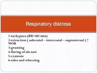
RESPIRATORY DISTRESS SYNDROME (RDS) • RDS is still probably the most common problem in the nursery, associated with premature delivery. The incidence and severity of RDS increases with gestational age at birth and is usually worse in male infants. • In fact, RDS is not a disease, but a syndrome that presents the foliowing early signs and symptoms: • Tachypnea>60 breath/min. • Respiratory effortwhich means: -expiratory grunting, nasal flaring, chest retraction, intercostal tirrage; use of accessory muscle of breathing; • Cyanosis:acrocyanosis or generalized cyanosis.Silverman scoremay quantify the respiratory effort and give a prognosis: -0 score indicates the absence of RDS and - >6 indicates a severe form of RDS.
RESPIRATORY DISTRESS SYNDROME (RDS) • In respiratory distress syndrome auscultation may find rales, ronchus, poor air exchange. • Chest radiographs are characterized by: • Atelectasis; • Air bronchograms; • Diffuse reticular-granular infiltrates often progressing to sever bilateral opacity characterized by therm whiteout. • Radiographs pattern in RDS is variable and may not reflect the degree of respiratory compromise. • Morphopathological findings in the course of RDS includes: • Atelectasis; • Pulmonary edema; • Pulmonary vascular congestion; • Pulmonary hemorrhage; • Evidence of direct injury to the respiratory epithelium;
RESPIRATORY DISTRESS SYNDROME (RDS) Avery first demonstrates the paucity of alveolar surfactant in thelungs of infants dying of RDS. Lack of pulmonary surfactant leads to: • Progressive atelectasis; • Alteration in ventilation perfosion ratio(V/P ratio); • Large areas of lung not perfused ; • Large right-to-left shunt of blood; • Pulmonary capillary blood flow decreased; • Alveolar ventilation decreased; • Lung volume reduced; Many of these pathologic findings may be observed in diseases which presents signs and symptoms of respiratory distress syndrome.This diseases may be divided in pulmonary and extrapulmonary disorders, both with RDS.
RESPIRATORY DISTRESS SYNDROME (RDS) • I.Pulmonary disorders with RDS A.Common • Hyaline membrane disease (HMD) • Transient tachypnea (TTN) • Meconium aspiration syndrome (MAS) • Pulmonary hypertension • Pneumonia B.Less common • Pulmonary hemorrhage • Pneumothorax • Imature lung syndrome C.Rare • Upper airway obstruction(e.g. choanal atresia) • Pulmonary hypoplasia • Space occuping lession(e.g.diaphragmatic hernia, lung cysts) • Rib cage anomalies
RESPIRATORY DISTRESS SYNDROME (RDS) II.Extrapulmonarv disorders with RDS(1) A.Heart 1. Congenital heart diseases B.Blood 1. Acute blood loss 2.Hypovolemia 3. Anemia 4.Polycytemia C.Metabolic 1. Acidosis 2.Hypoglicemia 3.Hypomagnesemia 4.Hypo/hypernatremia 5.Hypocalcemia
RESPIRATORY DISTRESS SYNDROME (RDS) II.Extrapulmonarv disorders with RDS (2) D.Septicaemia E.Brain • Hemorrhage • Edema • Trauma-mechanic • Trauma- hypoxic-ischemic encephalophaty • Congenital malformation of CNS F.Surgical causes • Intestinal obstructions. • Diphragmatic hernia G.Drugs administerred to the mother/newborn
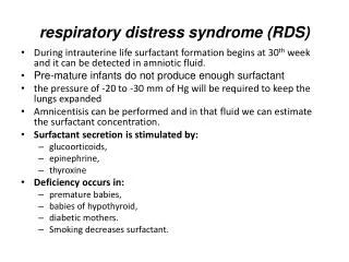
RESPIRATORY DISTRESS SYNDROME (RDS)-HMD HYALINE MEMBRANE DISEASE • HMD, also called respiratory distress syndrome is a pulmonary disease of the newborn with immature lungs and surfactant deficiency. • Incidence-the incidence of HMD is reverse proportional with premature deliveries. • 20% at newborn of 32-34 w.g., • 40% at 30-32 w.g. and • 0,5-1% at term babies
RDS.HMD • Factors increasing the risk of HMD: • Prematurity-HMD is very common in infants under 32 w.g. and most invariable under 28 w.g. • Male sex-androgens induces a delayed maturation of lecithin(a compound of surfactant). • White race (reasons unknown). • Caesarian section delivery - about 1/3 of fetal lung fluid is removed by squeezing the baby chest during vaginal delivery. • Asphyxia-during this state, the lung perfusion falls, the enzymatic activity is reduced and surfactant synthesis falls. • Maternal diabetes-insulin delayed the maturation of surfactant. • Familial predisposition. • Second born twin. • Hypothermia-surfactant function is defective in cold babies.
RDS.HMD • Factors decreasing the risk of HMD: • Pregnancy induced hypertension • Chronic maternal hypertension • Subacute placental abrubtio • Maternal narcotic addiction • Premature rupture of membranes • Prenatal corticosteroides
RDS.HMD Pathophysiology: • The major cause of HMD is surfactant deficiency, which results when the rate of use the surfactant exceeds the rate of production. Surfactant is produced in type II alveolar epithelial cells. Corticosteroids -endogenous or exogenous-and thyroid hormone increase the rate of synthesis and insulin decrease the synthesis of surfactant. • Surfactant is mainly composed of phospholipids-90%-, with small amounts of neutral fat, cholesterol and proteins. Their main functions are: • To lower surface tension • Anti-atelectatic action • Decrease ventilator effort • Role in host defense mechanism in the lung; The major cause of HMD is surfactant deficiency
RDS – risk factors PERINATAL ASPHIXIA PREMATURITY ACIDOSIS Surfactant deficiency ATELECTASIS Alveolar hypoventilation Mismatch V/P HYPOXEMIA RESPIRATORY ACIDOSIS Pulmonary VSC Injury to epit.cells,edema,vasc.congestion,hemorrhage Organization of hyaline mb
RESPIRATORY DISTRESS SYNDROME (RDS) Clinical presentation History Preterm infant with/without history of asphyxia in the perinatal period. It presents with some respiratory difficulties at birth that become progressively more severe. Physical examination • Cyanosis in room air; • Nasal flaring; • Tachypnea; • Grunting on expiration; • Retraction of chest wall. The infant grunts to prolong expiration and this mechanism actually improves alveolar ventilation. The retractions occur and increase as the infant is forced to develop high transpulmonary pressure to reinflate atelectatic air spaces.
RDS Paraclinical exams • X-ray-an anteroposterior chest x-ray film should be obtained for all infants with respiratory distress of any duration. The typical radiographic finding is a uniform reticulogranular pattern, referred to as a ground glass appearance, accompanied by air bronchograms.During the clinical course, sequential x-ray films may reveal air leaks secondary to ventilatory intervention as well as the onset of changes compatible with BPD. • Echocardiography-is a valuable diagnostic tool in the evaluation of an infant with hypoxemia and respiratory distress. Significant congenital heart disease can be excluded by this technique. • Transfontanelar US - proves neurological dammage.
RDS Laboratory studies: • Blood gas sampling is essential in the management of HMD. Hypoxemia is the hallmark of the disease and can be used to assess its severity; hypercarbia may be often seen and also acidosis-initial respiratory and then metabolic or mix acidosis.Although there is no consensus, most neonatologists agree that: • Pa02 of 50-70 mmHg and • PaC02 of 45-60 mmHg are acceptable; • most would maintain the pH at or above 7,25 and • the arterial 02 saturation at 88-95%.
RDS Laboratory studies: • Hemoglobin or hematocrit measurement; • Serum glucose level must be monitories for the risk of hypoglycemia; • Septic workup-CBC count, platelet count, blood culture, amniotic fluid culture and urine culture should be considered for each infant with a diagnosis of HMD • Serum electrolyte levels should be followed every 12-24 h for management of parenteral fluids; • Serum calcium level should be followed since hypocalcemia is common in sick,nonfed,preterm,or asphyxiated infants • Explore the renal function for severe asphyxiated infants-urea and creatinine level.
RDS Positive diagnosis: • Clinical presentation(history and physical examination) • Chest x-ray study • Laboratory studies
RDS Differential diagnosis • TTNB-may be present with a similar pictureas HMD, but they present a gradual improvement, with a lower requirement of 02 .x-ray will be very helpful; • Infection-bacterial pneumonia-very difficult to exclude in such babies, so we must routinely treat with antibiotics all infants with HMD, pending negative cultures; • Aspiration pneumonia, from either meconium or aspiration fluid, may present like HMD but is very unusual in prem. infants and demonstrates a different RX-appearance; • Congenital heart disease-echocardiography; • Pulmonary edema from primary cardiovascular anomalies, especially, patent ductus arteriosus (PDA), may mimic HMD or complicate it's course; • Upper airway obstruction–stridor present, gases normal;
RDS.HMD Management A.Prevention: • Avoid prematures deliveries and correct follow-up of risk pregnancies; • Prevention of preterm labor,using both tocolytic agents and corticosteroids to induce lung maturation(celestone-betamethazone-4mg at every 6h with 48h before of delivery and dexamethazone-2 doses of 12 mg at 24 h before delivery). B.Respiratory support: • Oxygen therapy-by mask,hood,CPAP-nasal prong or nasopharyngeal CPAP. It works better on larger infants with mild to moderate disease and good respiratory effort. • Endotracheal intubation and mechanicalventilation are the mainstays of therapy for infants with RDS in whom apnea and hypoxemia with respiratory acidosis develops.
RDS.HMD C.Fluid and nutriţional support: • to provide energy, prevent hypoglycemia and assure the metabolic basal needs– this is done by parenteral nutrition, sometimes for an extended period. The specific needs of preterm infants are becoming better understood and the nutrient preparation available reflects this understanding.
RDS D.Antibiotic therapy-antibiotics that cover the most common neonatal infections are usually begun initially, because of dificulty in distinguish HMD from bacterial pneumonia and because these premature babies had a high risk for infections. E.Surfactant replacement-is now typically used in the treatment of HMD. Surfactant administration early in the course of HMD restores pulmonary function and improves ventilation to open, poorly ventilated lung units and increases their alveolar P02.As result, hypoxic vasoconstriction is relieved, the right-to-left shunt decreases and overall arterial oxygenation improves. Three types of surfactant are available: Survanta and Curosurf - modified natural surfactants, from purified extracts of animal lung- and Exosurf- a completely synthetic surfactant, made of phospholipids.
RDS Outcome and complications: Many infants who have HMD recover without sequelae. Although mortality increases with decreased gestational age, administration of antenatal corticosteroids to the mother and surfactant to the newborn can significantly improve survival. Immediate complications are: • Pulmonary air leaks, such as pneumothorax, pneumomediastinum, pneumopericardum; • Intraventricular hemorrhage; • Patent ductus arteriosus; • Infections such as pneumonia, septicemia; • Tracheal stenosis;
RDS Outcome and complications: Late complications are: • Bronchopulmonary dysplasia; • ROP; • Neurological sequelae.
TRANSIENT TACHYPNEA OF THE NEWBORNT.T.N. Definition TTN is also known as wet lung or type II RDS. It is a benign disease of near-term, term, or large premature infants who have respiratory distress shortly after delivery that usually resolves within 3 days. Incidence-1-2% of all newborns Risk factors: • Elective cesarean section delivery. • Male sex. • Macrosomia. • Excessive maternal sedation. • Prolonged labor. • Birth asphyxia. • Fluid overload to the mother, especially with oxytocin infusion. • Delayed clamping of the umbilical cord (optimal time is 45 sec). • Breech delivery. • Fetal polycythemia. • Infant of a diabetic mother.
TRANSIENT TACHYPNEA OF THE NEWBORN Pathophysiology: At term, the fetal lung contains about 20-ml/kg/body weight of fluid. This fluid is contained in airways and air sacs and is secreted by fetal lung. Clearance of the fetal ling liquid begins before birth-during the last few days of gestation-and during labor. TTN is thought to occur because delayed resorption of fetal lung fluid from the pulmonary lymphatic system. The increased volume caused a reduction in lung compliance and increased airway resistance. This result in tachypnea and retractions. Infants delivered by elective cesarean section are at the risk because of lack of the normal vaginal thoracic squeeze, which force lung fluid out. One hypothesis is that TTN may represent a mild surfactant deficiency in these infants.
TRANSIENT TACHYPNEA OF THE NEWBORN Clinical presentation: • The infant with TTN usually has mild to moderate disease characterized by: • Tachypnea>80 breath/min within 2 to 6 hours after delivery; Very rare we meet: • Cyanosis; • These signs persist for 12 to 72 hours, according to natural evolution of the disease, or to the provided management.
TRANSIENT TACHYPNEA OF THE NEWBORN Diagnosis -anamnesis and clinical exam; -laboratory studies: -arterial blood gas on room air will show some degree of hypoxia; -complete blood cell count is normal in TTN but should be obtained if considering an infectious process; Radiological studies-the typical findings in TTN are: - hyperexpansion of the lung, a hallmark of TTN; - prominent perihilar streaking-secondary to engorgement of periarterial lymphatic. - mild to moderate enlarged heart; - fluid in the minor fissure.
TRANSIENT TACHYPNEA OF THE NEWBORN Differential diagnosis • Bacterial pneumonia-sometimes is difficult to distinguish from TTN.We routinely treat all infants with abnormal X-ray and a persistent O2 requirement with antibiotics with broad spectrum until cultures are negatives. • Heart disease-the 100% 02 test should be done to rule out heart disease. • HMD-the infant will normally be premature or have some reason for delayed lung maturation, such as maternal diabetes. The chest X- ray will be helpful in these cases. • Metabolic disorders- infants with hypothermia, hyperthermia, or hypoglycemia may all have tachypnea.
TRANSIENT TACHYPNEA OF THE NEWBORN Management • TTN is a self-limited disease that usually resolves completely within 48-72 hours. Initial management consists in providing adequate oxygenation to keep arterial oxygenation greater than 90%. Intravenous antibiotics should be administered until infection has been excluded. Modest fluid restriction of 60 ml/kg/day LV. is appropriate while assessing the need for supplemental oxygen. • Administration of diuretics does not alter the course of the disease. Prognosis TTN is a benign disease with no risk for further pulmonary dysfunction.
MECONIUM ASPIRATION SYNDROME Definition: Meconium is the first intestinal discharge of the newborn infant and is composed of epithelial cells, fetal hair, mucus, and bile. Intrauterine stress may cause in utero passage of meconium into the amniotic fluid. The fetus may aspirate the meconium-stained amniotic when hypoxia and hypercapnia stimulate fetal gasping or deep breathing movements. The presence of the meconium in the trachea may cause airway obstruction as well an inflamatory response, resulting in severe respiratory distress. The presence of meconium in amniotic fluid is a warning sign of fetal distress, but is not a sensitive marker of fetal distress. Incidence: The incidence of meconium-stained amniotic fluid varies from 8 to 15% of all deliveries. M.A.S. primarily affects termand postmature infants.
MECONIUM ASPIRATION SYNDROME Risk factors: -the following factors have been associated with an increased risk of meconium passage and subsequent aspiration: • Postterm pregnancy; • Preeclamsia-eclamsia; • Maternal hypertension; • Maternal diabetes mellitus; • Abnormal fetal heart rate; • Intrauterine growth retardation (IUGR); • Oligohydramnios;
MECONIUM ASPIRATION SYNDROME Pathophysiology: Meconium first appears in fetal ileum between 10 and 16 weeks of gestation. Control of fetal meconium passage is dependent on hormonal and parasympathetic neural maturation. After intrauterine passage of meconium, deep irregular respiration or gasping, either in utero or during labor or delivery, can cause aspiration of the meconium stained amniotic fluid. Thick, meconium stained amniotic fluid can result in acute upper airway obstruction. As the aspirate meconium progresses distally, total or parţial airway obstruction may occur. In areas of total obstruction, atelectasis developps but in areas of partial obstruction, a ball-valve phenomenon occurs, resulting in air trapping and hyperextension. With distal progression of meconium, interstitial and chemical pneumonitis develops, with resulting bronchiolar edema and narrowing of the small airways. Meconium at the alveolar level may inactivate surfactant.
MECONIUM ASPIRATION SYNDROME Intrauterine hypoxia MECONIUM ASPIRATION Meconium stained amniotic fluid Upper airway obstr. Lower airway obstr. Diffuse particle spread Chemical pneumonitis complete partial ACUTE HYPOXIA ATELECTASIS Ball effect INFECTION Mismatch V/P Obstructive Emphysema Inhibition Surfactant synthesis PPH Low compl Hypoxia,Hypercapnia,Acidosis metab.& resp
MECONIUM ASPIRATION SYNDROME Clinical presentation The clinical aspect of an infant with M.A.S. is variable. Symptoms depend on the severity of the hypoxic insult and the amount and viscosity of the meconium aspirated. The meconium present in amniotic fluid varies in appearance and viscosity, ranging from thin, green-stained fluid to a thick "pea soup" consistency. The infants with M.A.S. often exhibit signs of postmaturity: long nails and peeling yellow-or green-stained skin. Early meconium aspiration syndrome is characterized by airway obstruction. Large amounts of thick meconium, if not removed, can result in acute large airway obstruction. These infants may be apneic or have gasping respiration, cyanosis, and poor air exchange. Endotracheal suctioning must rapidly clear the airway.
MECONIUM ASPIRATION SYNDROME Clinical presentation: The infants who aspirated meconium into distal airways but do not have total airway obstruction manifest signs of respiratory distress: tachypnea, nasal flaring, intercostal retraction, increased anteroposterior diametre of the chest and cyanosis. Some infants may have delayed symptoms, with only mild initial respiratory distress, which becomes more severe hours after delivery as atelectasis and chemical pneumonitis develop.
MECONIUM ASPIRATION SYNDROME Diagnosis: • Chest X-ray-reveals hyperinflation the lung fields a flattened diaphragms. There are coarse, irregular patchy infiltrates alternating with areas of over expansion. Pneumothorax and pneumomediastinum may be present. The severity of X-ray may not always correlate with the clinical disease. • Laboratory studies-arterial blood gas levels characteristically reveal hypoxemia. Hyperventilation may result in respiratory alkalosis in mild cases, but infants with severe disease usually manifest respiratory acidosis due to airway obstruction, atelectasis and pneumonitis,. If the patient has suffered perinatal asphyxia, combined respiratory and metabolic acidosis is present.
MECONIUM ASPIRATION SYNDROME Management: A.Prenatal management • identification of high risk pregnancies; • monitoring-during labor careful observation and monitoring should be performed. B.Delivery room management • The severity of meconium aspiration can be markedly decreased by early removal of aspirated tracheal meconium. Those infants who are depressed or have thick, meconium stained fluid should be intubated after their trachea suctioned using a meconium trap aspirator. • The approach to infants who are vigorous or who have thin or moderately stained amniotic fluid remains controversial.
MECONIUM ASPIRATION SYNDROME Management of the newborn with M.A.S. Infants with meconium below their trachea are at risk for pulmonary hypertension, air leak syndromes, and pneumonitis and must be observed closely for signs of respiratory distress. 1 .Respiratory management a. Pulmonary toilet-if suctioning the trachea does not result in clearing ofsecretions, it may be advisable to leave an endotracheal tube in place,_insymptomatic infants for pulmonary toilet. Chest physiotherapy every 30’-1 hour tolerated, will aid in clearing the airway. b. ABG level-should be obtained for assessing ventilatorcompromise and supplemental oxygen requirements. c. Oxygen monitoring-a pulsoximeter will provide important informationregarding the severity of the child's respiratory status and will also assistin preventing hypoxemia.
MECONIUM ASPIRATION SYNDROME d.Chest X-ray films-should be obtained after delivery. A chest X-raymayalso help determine which patients will develop respiratory distress.However, the radiograph often poorly correlates with clinical presentation. e. Antibiotic coverage-meconium inhibits the normally bacteriostaticquality of amniotic fluid. Since it is difficult to differentiate meconiumaspiration from pneumonia radiographically, infants with infiltrates onchest X-ray should be started on broad-spectrum antibiotics afterappropriate cultures have been obtained. f. Supplemental oxygen-a major goal is to preventepisodes of alveolarhypoxia leading to hypoxic pulmonary vasoconstriction and thedevelopment of persistent pulmonary hypertension of the newborn. Forthat purpose, supplemental oxygen is provided "generously", such thatarterial oxygen tension is maintained at least in the range of 80-90 mmHg.
MECONIUM ASPIRATION SYNDROME g.Mechanical ventilation-patient with severe disease that are in respiratoryfailure with hypercarbia andpersistent hypoxemia require mechanicalventilation. Ventilation must be adjusted to the individual patient. Thesepatients typically require higher inspiratory pressures and faster rates thanpatient with HMD does.Those infants who do not respond to conventional ventilation should be given high frequency ventilation. h.Surfactant administration-infants with severe M.A.S. may benefit fromsurfactant therapy.
MECONIUM ASPIRATION SYNDROME 2.Cardiovascular management-persistent pulmonary hyperten- sion is frequently associated with M.A.S.To minimize this risk, aggressive resuscitation and stabilization are essential. • These infants may also require specific therapy for hypotension and poor cardiac output, including including temporary support with colloid infusion or cardiotonic medication such dopamine. 3.General management-infants that have aspirated meconium and require resuscitation often develop metabolic abnormalities such as hypoxia, acidosis, hypoglycemia and hypocalcemia. • Because these patients may have suffered perinatal asphyxia, surveillance for any end-organ damage is essential.
MECONIUM ASPIRATION SYNDROME Complications: • Because these patients may suffer perinatal asphyxia, hypoxic ischemic encephalophathy may be severe consequences. • Pneumothorax and airleaks are the most common complication occurring in 15-20%. • Pulmonary hypertension usually responds promptly to pentoxiphylin administration. • The risk if infection in these babies is similar to that in all babies who are ventilated.