Download
1 / 6
60 likes | 88 Views
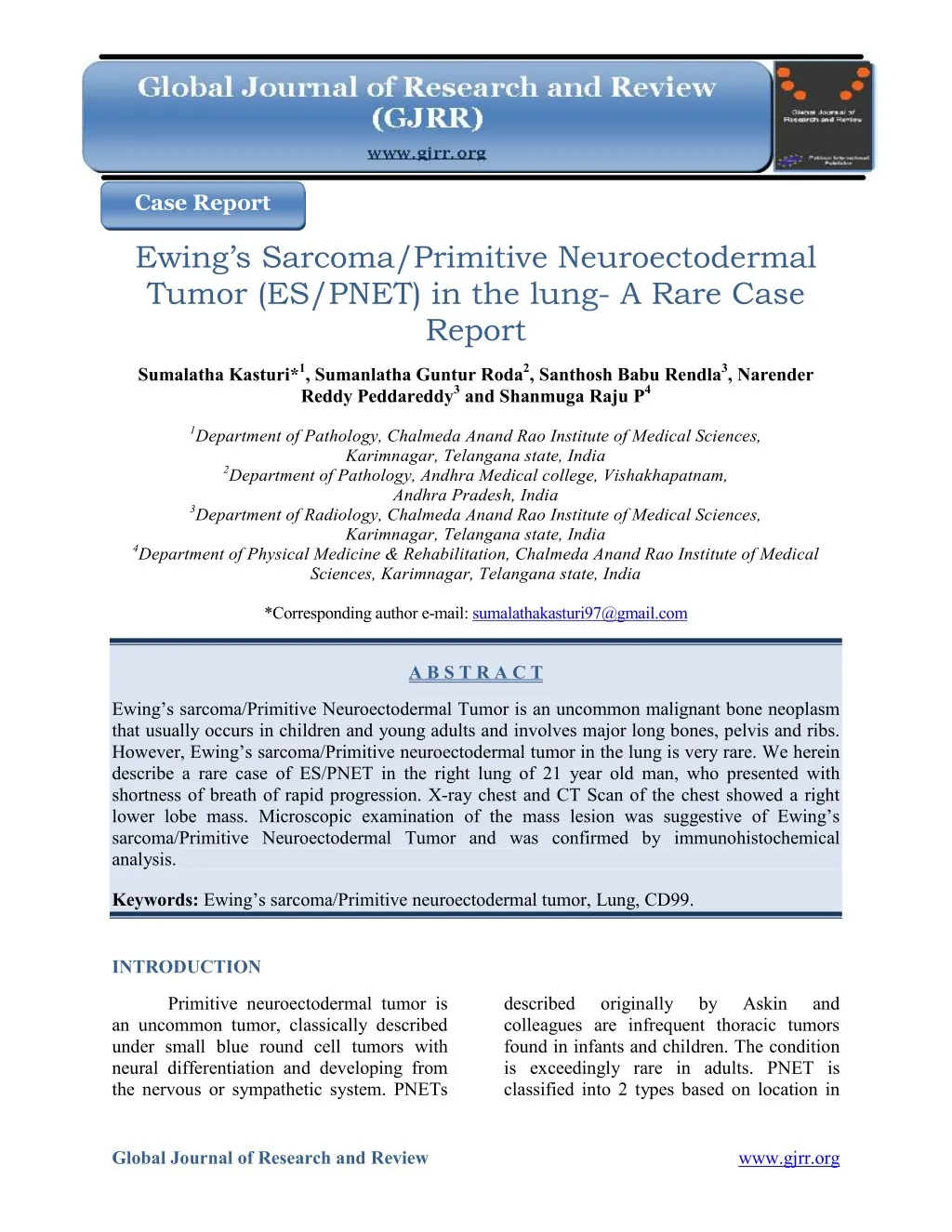
Ewing’s sarcoma/Primitive Neuroectodermal Tumor is an uncommon malignant bone neoplasm that usually occurs in children and young adults and involves major long bones, pelvis and ribs. However, Ewing’s sarcoma/Primitive neuroectodermal tumor in the lung is very rare. We herein describe a rare case of ES/PNET in the right lung of 21 year old man, who presented with shortness of breath of rapid progression. X-ray chest and CT Scan of the chest showed a right lower lobe mass. Microscopic examination of the mass lesion was suggestive of Ewing’s sarcoma/Primitive Neuroectodermal Tumor and was confirmed by immunohistochemical analysis

E N D
Case Report Ewing’s Sarcoma/Primitive Neuroectodermal Tumor (ES/PNET) in the lung- A Rare Case Report Sumalatha Kasturi*1, Sumanlatha Guntur Roda2, Santhosh Babu Rendla3, Narender Reddy Peddareddy3 and Shanmuga Raju P4 1Department of Pathology, Chalmeda Anand Rao Institute of Medical Sciences, Karimnagar, Telangana state, India 2Department of Pathology, Andhra Medical college, Vishakhapatnam, Andhra Pradesh, India 3Department of Radiology, Chalmeda Anand Rao Institute of Medical Sciences, Karimnagar, Telangana state, India 4Department of Physical Medicine & Rehabilitation, Chalmeda Anand Rao Institute of Medical Sciences, Karimnagar, Telangana state, India *Corresponding author e-mail: sumalathakasturi97@gmail.com A B S T R A C T Ewing’s sarcoma/Primitive Neuroectodermal Tumor is an uncommon malignant bone neoplasm that usually occurs in children and young adults and involves major long bones, pelvis and ribs. However, Ewing’s sarcoma/Primitive neuroectodermal tumor in the lung is very rare. We herein describe a rare case of ES/PNET in the right lung of 21 year old man, who presented with shortness of breath of rapid progression. X-ray chest and CT Scan of the chest showed a right lower lobe mass. Microscopic examination of the mass lesion was suggestive of Ewing’s sarcoma/Primitive Neuroectodermal Tumor and was confirmed by immunohistochemical analysis. Keywords: Ewing’s sarcoma/Primitive neuroectodermal tumor, Lung, CD99. INTRODUCTION Primitive neuroectodermal tumor is an uncommon tumor, classically described under small blue round cell tumors with neural differentiation and developing from the nervous or sympathetic system. PNETs described colleagues are infrequent thoracic tumors found in infants and children. The condition is exceedingly rare in adults. PNET is classified into 2 types based on location in originally by Askin and Global Journal of Research and Review www.gjrr.org
Kasturi et al__________________________________________________ISSN 2393-8854 the body: Peripheral PNET (ES) and CNS PNET. Peripheral PNET is now thought to be identical to Ewing’s Sarcoma, based on evidence that indicates that both ES and PNET have a similar neural phenotype and, because they share an identical chromosome translocation1,2. Primitive neuroectodermal tumors are tumors that demonstrate neural differentiation by light microscopy, IHC, or electron microscopy, where as Ewing’s sarcomas are undifferentiated by these analysis. PNET of the CNS generally refers to supratentorial PNETs2. To the best of our knowledge, only eight cases of primary pulmonary ES/PNET have been reported in the literature. We present here such a rare case of Ewing’s Neuroectodermal Tumor in the lung of young adult male. CASE REPORT eosinophilic cytoplasm and indistinct cell borders (figure-4, 5). Some of the cells had vacuolated cytoplasm. At this point, small blue round cell tumor was suspected. The sections were then immunohistochemistry for CK, LCA, TTF-1 and CD99. IHC staining for CD99 (figure-6) showed positive result and there was negative staining for CK, LCA and TTF-1. Based on histomorphology and IHC the case was diagnosed as Ewing’s Sarcoma/ Primitive Neuroectodermal Tumor in the lung. DISCUSSION subjected to Ewing’s Sarcoma and extraskeletal Ewing's sarcoma were first described by James Ewing and Tefft3,4. Since then these two conditions have histologically with other small blue round cell tumors, including PNET3,4, but recent immunoperoxidase and cytogenic studies showed that PNET and ES belongs to the same group with varying degrees of neural differentiation, and they were categorized into a group known as the Ewing family of tumors14. The ES/PNET family is a rare malignant tumor that shares common histological features of small primitive round cells. It most often arises in the soft tissues and bones, but has rarely been reported on other sites, such as the ovary, testis, uterus, kidney, pancreas, palate, myocardium, and morphological features of intrapulmonary tumors are similar to those of the ES/PNET in a variety of other locations. The tumors are diagnosed using several modalities such as light microscopy, immunohistochemistry, and cytogeneteics6. Immunohistochemical staining is positive for neuron-specific enolase (60%), S-100 protein (50%), and CD99 marker (90%), as well as negative findings for leukocyte common antigen, epithelial membrane antigen, cytokeratin9. In this case, positive sarcoma/Primitive been confused A young adult of 21 years complained of shortness of breath, cough and pain in chest since 20 days. He was a non-smoker with no history of exposure to any occupational or inorganic dusts. There was no clubbing and lymphadenopathy on general examination. The respiratory system evaluation revealed signs of mass in the right hemithorax. Complete blood counts and biochemical investigations were within normal limits. X-ray chest showed a mass in the right lower lobe (figure-1). CECT of the thorax showed a large heterogenous lower lobe mass, in the right hemithorax (figure- 2). There was no involvement of mediastinal lymph nodes. Right lower lobectomy was done and sent for examination. Grossly, cut section of the tumor was gray white with large areas of hemorrhage and Histologically, the tumor showed closely packed uniform round cell sheets with intervening delicate fibrovascularstroma. The cells show small round to ovoid nucleus with fine powdery chromatin and scant pale lung5-8,14). The histopathological necrosis (figure-3). GJRR[2][2][2015] 058-063
Kasturi et al__________________________________________________ISSN 2393-8854 4.Tefft M, Vawter GF, Mitus A. Paravertebral round celltumors in children. Radiology. 1969; 92:1501–1509. 5.Kang MS, Yoon HK, Choi JB, Eum JW. Extraskeletal Ewing's sarcoma of the hard palate. J Korean Med Sci. 2005; 20:687– 690. 6.Tsuji S, Hisaoka M, Morimitsu Y, Hashimoto H, Jimi A, Watanabe J, Eguchi H, Kaneko Y. neuroectodermal tumour of the lung: report of two cases. Histopathology. 1998; 33: 369–374. 7.Charney DA, Charney JM, Ghali VS, Teplitz C. Primitive neuroectodermal tumor of the myocardium: a case report, review of the literature, immunohistochemical, and ultrastructural study. Hum 27:1365–1369. 8.Koo HL, Jun SY, Choi G, Ro JY, Ahn H, Cho KJ. Primary Primitive neuroectodermal tumor of the kidney: report of two cases. Korean J Pathol. 2003; 37:145–149. 9.Christie DR, Bilous AM, Carr PJ. Diagnostic difficulties in extraosseous Ewing's sarcoma: a proposal for diagnostic criteria. Australas Radiol. 1997; 41:22–28. 10.Kahn AG, Avagnina A, Nazar J, Elsner B. Primitive neuroectodermal tumor of the lung. Arch Pathol Lab Med. 2001; 125:397– 399. 11.Imamura F, Funakoshi T, Nakamura S, Mano M, Kodama K, Horai T. Primary primitive neuroectodermal tumor of the lung: report of two cases. Lung Cancer. 2000; 27:55–60. 12.Cotterill SJ, Ahrens S, Paulussen M, Jurgens HF, Voute PA, Gadner H, Craft AW. Prognostic factors in Ewing's tumor of bone: analysis of 975 patients from the European Intergroup Cooperative Ewing's Sarcoma Study Group. J. 18:3108–311. 13.13.Eroglu A, Kurkcuoglu IC, Karaoglanoglu N, Alper F, Gundogdu C. Extraskeletal Ewing sarcoma of the diaphragm presenting with hemothorax. Ann Thorac Surg. 2004; 78:715–717. 14.14. Danner DB, Hruban RH, Pitt HA, Hayashi R, Griffin CA, Perlman EJ. Primitive neuroectodermal tumor arising in staining for CD99 was observed confirming the diagnosis. The CT-scan finding of ES/PNET reported is a heterogeneously enhancing mass11.In our case also heterogeneously enhancing opacity was observed. The treatment for ES/PNET is aggressive with the most effective treatment being a surgical resection with combination chemotherapy and high-dose radiation therapy. When resectable, the surgical excision must be wide12,13. Right lower lobectomy was performed and combination chemotherapy was administered. As multidisciplinary physiotherapy and rehabilitation were given to achieve optimal survival and better quality of life. CONCLUSION Peripheral primitive subsequently part approach, a team of a Pathol. 1996; Although, the ES/PNET in the lung is very rare soft tissue tumor, it should be considered in the differential diagnosis of a lung mass. The diagnosis is difficult on light microscopy alone immunohistochemistry for confirmation. These tumors present at a younger age, and are very aggressive. They usually have a poor prognosis despite treatment. REFERENCES and relies on 1.Maureen J. O’ Sullivan, Elizabeth J Perlman, Jaime Furman et al. Visceral primitive peripheral tumors: A Clinicopathologic and molecular study. Hum Pathol. 1997; 44; 1799-1802. 2.Kumar, Vinay, Fausto, Nelso, Abbas, Abul. Robbins &Cotran Pathologic Basis of Disease, 8th ed. Saunders. Page 1301. 3.Ewing J. Classics in oncology. Diffuse endothelioma of bone. Proceedings of the New York Pathological Society, 1921. CA Cancer J Clin. 1972; 22:95–98. neuroectodermal Clin. Oncol. 2000; GJRR[2][2][2015] 058-063
Kasturi et al__________________________________________________ISSN 2393-8854 the pancreas. Mod Pathol. 1994; 7: 200-204. [PubMed]. 15.15. Shin JH, Leett K, Rhim SC, Chok J, Choi CG, Suh DC. Spinal epidural extraskeletal Ewing sarcoma: MR findings in two cases. AJNR Am J Neuro Radiol. 2001; 22:795-798. [PubMed]. Figure 1. Chest radiograph showing mass lesion in the right lower lobe with right pleural effusion Figure 2. CT Scan thorax showing heterogenously enhancing mass lesion in the right lower lobe GJRR[2][2][2015] 058-063
Kasturi et al__________________________________________________ISSN 2393-8854 Figure 3. Gross picture showing friable and haemorrhagic lesion involving entire lower lobe Figure 4. Microscopy showing closely packed tumor cells in sheets separated by thin fibrovascular septae. H&E, x100 GJRR[2][2][2015] 058-063
Kasturi et al__________________________________________________ISSN 2393-8854 Figure 5. High power showing uniform round to oval cells with powdery chromatin, H&E, x400 Figure 6. Immunohistochemically tumor cells showing membrane positivity for CD99, x400 GJRR[2][2][2015] 058-063