Download
1 / 31
310 likes | 431 Views
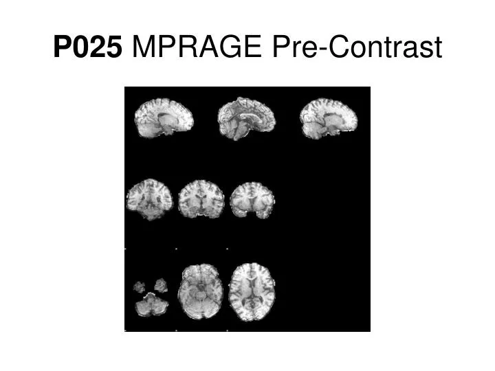
P025 MPRAGE Pre-Contrast. P025 MPRAGE w/ Z-Score < -4. Notes. This was accomplished by doing an inverse nonlinear warp from MNI to the SPGR FA 18, then to the MPRAGE space with a linear transform (ideally these transforms should be combined)

E N D
Notes • This was accomplished by doing an inverse nonlinear warp from MNI to the SPGR FA 18, then to the MPRAGE space with a linear transform (ideally these transforms should be combined) • Images are larger than shown, zoom in for more detail • Not all lesions are found by the z-score thresholding but a significant amount are • Large volumes of white matter are found to be significantly demyelinated: perhaps DAWM or actually revealing the “invisible disease”? • This was one the patients with a larger amount of low MWF volume, EDSS 5.0 SPMS
MPRAGE • Same slices as shown in the z-score slides • Lesions of interest circled
SPGR 3 class-CSF • Generally good CSF segmentation • Does not capture most lesions
SPGR 3 class-GM • Includes some or all of lesions
SPGR 3 class-WM • Matches nicely with the MPRAGE scan • Partially includes lesions
SPGR 4 class-CSF • Generally worse than 3 class CSF
SPGR 4 class-GM • More conservative estimate of GM, much fewer lesions included
SPGR 4 class-More GM • Deep GM • Includes many of the lesions
SPGR 4 class-WM • Does well at excluding most focal lesions but appears to be some partially including some • More conservative
SPGR-FLAIR 3 class-CSF • Grabs most lesions • Unfortunately mis-classifies GM too
SPGR-FLAIR 3 class-GM • Includes many regions previously seen as WM
SPGR-FLAIR 3 class-WM • Good exclusion of lesions identified by z-score • Does poorly at WM and GM segmentation, misses some WM
SPGR-FLAIR 4 class-CSF • Generally worse than 3 class CSF also • Chokes back mask too far
SPGR-FLAIR 4 class-GM • Still misidentifies a lot of WM as GM • Catches edges of our lesions of interest
SPGR-FLAIR 4 class-More GM • Again mis-includes a lot of WM
SPGR-FLAIR 4 class-WM • Avoids lesions but also misses a lot of regular WM since those are misidentified as GM
SPGR-T2-PD 3 class-CSF • Decent at outside brain CSF, though catches some WM • Does not get any CSF inside brain
SPGR-T2-PD 3 class-GM • Captures our lesions of interest • Gets ventricle CSF • Not as good as SPGR 3 class
SPGR-T2-PD 3 class-WM • Overly greedy WM • Misses focal lesions but gets GM
SPGR-T2-PD 4 class-CSF • Same problems as with SPGR-T2-PD CSF segmentation
SPGR-T2-PD 4 class-GM • GM + inner CSF • Catches darker lesions • Pretty poor, also gets WM
SPGR-T2-PD 4 class-More GM • Catches some lesions but overall pretty garbagey • Doesn’t really correspond to a distinct tissue class
SPGR-T2-PD 4 class-WM • A conservative estimate • Circled region shows possible inclusion of GM and missing of brain stem
Best CSF • SPGR 3 class (includes some lesions) • SPGR-FLAIR 3 class (includes all lesions) • SPGR-T2-PD 3 class (useful for out of brain CSF, does not include lesions)
Best WM • SPGR 3 class (best anatomically) • SPGR 4 class (conservative) • SPGR-FLAIR 4 class (more lesion exclusion)
Best GM • SPGR 3 class (GM+lesions) • SPGR 4 class (deep GM+lesions)
Best Lesion • SPGR 3 class (GM+lesions) • SPGR-FLAIR 3 class (CSF missing some lesions)
Coming Soon • Segmentation with a priori maps • Ideas about how to combine maps to produce NAWM, NAGM, and lesion only masks