Download
1 / 42
470 likes | 633 Views
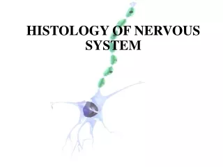
Histology of Nervous Tissue. Cells of nervous tissue. Neurons and supporting cells. Neurons Functional units of the nervous system - relay sensory (afferent) stimuli from the internal and external environments - process, store and integrate information

E N D
Cells of nervous tissue Neurons and supporting cells • Neurons • Functional units of the nervous system • - relay sensory (afferent) stimuli from the internal and external environments • - process, store and integrate information • - transmit information to other neurons, muscles and glands • Composed of a cell body, dendrites, axon and its terminal • arborization and synapses • Form complex and highly integrated circuits
Astrocyte Supportive cells Highly outnumber neurons CNS: Astrocytes Structural support Metabolic support Homeostasis Oligodendrocytes Insulation (myelin sheath) Microglia Phagocytosis Immunomodulation Ependymal cells PNS: Schwann cells Myelin Oligodendrocyte Microglia
Functional compartments of the neuron Cell body Dendrites Axon Synapses
Functional compartments of the neuron Cell body Dendrites Axon Synapses
Functional compartments of the neuron Cell body Dendrites Axon Synapses
Functional compartments of the neuron Cell body Dendrites Axon Synapses
Cell body (Soma, Perikaryon) Nucleus
Neuronal Cytoskeleton Neuro- filaments Neurofilaments (8-10 nm) Microtubules (20-25 nm) • Gives shape and support for the cell • (Neurofilaments) • Facilitates intracellular transport • (Microtubules) SER Microtubules Cross section Microtubules Neurofilaments Longitudinal section
Nissl bodies RER + polysomes Nissl RER Polysomes
Dendrites Form majority of receptive field Usually multiple and branched at acute angles May possess spines Transmit toward cell body (receptor potentials, graded) Organelles (similar to cell body)
Dendrites Form majority of receptive field Usually multiple and branched at acute angles May possess spines Transmit toward cell body (receptor potentials, graded) Organelles (similar to cell body)
Axon Usually singular Smaller caliber and often longer than dendrites Right angle branching Organelles Lacks Nissl High cytoskeleton content Originates at the axon hillock Transmits action potentials away from cell body Neurons Muscles and glands Terminal arborization Terminal buton Pre-synaptic element Initial segment of the axon
Classified according to post-synaptic structure Dendrite (Axodendritic) Cell body (Axosomatic) Axon (Axoaxonic) Muscle cell (neuromuscular junction)
Chemical synapses Pre-synaptic terminal (buton) Neurotransmitter (synaptic) vesicles Mitochondria Synaptic cleft Post-synaptic cell with postsynaptic density Terminal buton Synaptic vesicles Active zone on presynaptic membrane Transmitter release Synaptic cleft Post synaptic density Postsynaptic density Dendritic spine
Classification of neurons By shape and function Multipolar Most numerous and heterogeneous group Efferent or association Pseudounipolar Sensory (afferent) Bipolar Sensory, organs of special sense Developmental stage for all neurons
Motor (efferent) Association Types of neurons Sensory (afferent) Pseudounipolar Multipolar Bipolar Pseudounipolar
Segregation of axons and cell bodies in the nervous system In both CNS and PNS cell bodies are found in clusters and neuron processes travel in bundles. Such groupings are based on common function and/or common connections. CNS Gray matter - composed primarily of neuron cell bodies and supporting cells Cortex - neuronal cell bodies arranged in layers covering the outer layers of the brain, e.g., the cerebrum and cerebellum Nucleus - group of neuron cell bodies in the interior of the CNS White matter - regions of CNS rich in myelinated axons Appears white in fresh tissue due to presence of lipid-rich myelin Composed of tracts - bundles of axons that course together G G W W W G G W
Segregation of axons and cell bodies in the nervous system PNS Ganglia (singular = ganglion) - groups of cell bodies with a similar function Nerves (both cranial and spinal nerves) - bundles of axons coursing together Nerve Ganglion
(Multipolar) (Pseudounipolar) (Multipolar) Reflex arc
Supporting Cells • CNS • Astrocytes • Oligodendrocytes • Ependymal cells • Microglia • PNS • Schwann cells Neuroglial cells (neural ectoderm)
Astrocytes • Stellate morphology • Fibrous • Protoplasmic • Functions • Physical support • Nutritional support • Ion homeostasis • Transmitter uptake • Scar formation • Immune function Cell of origin for some brain tumors (astrocytomas)
Fibrous Cap Perivascular end foot Cap Protoplasmic
Oligodendrocytes • Interfascicular and satellite • Interfascicular oligodendrocytes form the myelin sheath in the CNS Interfascicular Satellite
Ependymal cells Line the ventricles and central canal of brain and spinal cord Cilia Microglial cells "Brain macrophages“ Derived from mesoderm
Schwann cells Satellite Schwann cells Surround cell bodies Ensheathing Schwann cells Surround axons
Myelinsheath • Segmental, multilamellar, lipid-rich wrapping of axons formed by the plasma membranes of: - Oligodendrocytes in CNS (interfascicular) - Schwann cells in PNS • Functions to increase speed of conduction (saltatory conduction) by insulating the axon
Internode Myelin sheath Node of Ranvier Internode: Single myelin segment Node of Ranvier: Specialized region of the axon between internodes, rich in sodium channels; site where depolarization occurs
Myelin Myelin Axon Axon Cross section of a myelinated axon
Myelinsheath CNS PNS • Oligodendrocytes in CNS Multiple internodes per cell Multiple axons per cell • Schwann cells in PNS One internode per cell One axon per cell Schwann cells Oligodendrocyte
Unmyelinated axons Schwann cell nucleus
Connective tissues in the peripheral and central nervous systems
Peripheral nervous system Endoneurium Perineurium Epineurium Ep P En
Central nervous system Meninges (meningitis) Dura mater Arachnoid membrane Pia mater
Dura mater Arachnoid membrane Pia mater Brain Dura mater Arachnoid membrane Pia mater Brain Blood vessel