Download
1 / 17
290 likes | 789 Views
Histology of Nervous Tissue. Neuroglia Neurons Myelination. Nervous Tissue: Support Cells. Support cells in the Central Nervous System (CNS) are grouped together as neuroglia Neuroglia literally means “nerve glue”

E N D
Histology of Nervous Tissue • Neuroglia • Neurons • Myelination
Nervous Tissue: Support Cells • Support cells in the Central Nervous System (CNS) are grouped together as neuroglia • Neuroglia literally means “nerve glue” • The function of neuroglia is to support, insulate, and protect the delicate neurons of the brain
Types of Neuroglia in CNS • Astrocytes • Star-shaped cells • Half of all brain tissue • Brace neurons; they keep the neurons in contact with their blood supply (capillaries) • Control the chemical environment of the brain by mopping up leaked ions
Types of Neuroglia in CNS • Microglia • Spiderlike phagocytes (white blood cells) • Dispose of debris like dead brains cells and bacteria
Types of Neuroglia in CNS • Ependymal cells • Lines the cavities of the brain and spinal cord • Circulate cerebrospinal fluid by beating their cilia • Cerebrospinal fluid fills the space the brain does not take up and forms a protective cushion around the brain and spinal chord
Types of Neuroglia in CNS • Oligodendrocytes • Wrap around nerve cells in the brain and spinal chord • Produce myelin sheaths • Myelin is a fatty, insulation covering the nerve cells; allows for the electrical signal to transmit faster (like wire coating)
Types of Neurolgia in PNS • Satellite cells • Protects neuron cell bodies which is where the nucleus of the cell if found • Schwann cells • Form myelin sheath in the peripheral nervous system (nerves of the body; not nerves of the CNS)
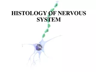
So what’s a Neuron? • Neurons = nerve cells • Cells specialized to transmit messages • Major regions of neurons • Cell body — nucleus and metabolic center of the cell (main part of nerve cell) • Processes — fibers that extend from the cell body • can be microscopic or up to 3-4 feet in length
Anatomy of a Neuron • Cell body • Nucleus • Large nucleolus • Processes outside the cell body • Dendrites — conduct impulses toward the cell body • Axons — conduct impulses away from the cell body
Anatomy of a Neuron • Axons end in axonal terminals • Axonal terminals contain small sacs with neurotransmitters (chemicals) • Axonal terminals are separated from the next neuron by a gap (they never really touch) • Synaptic cleft — gap (space) between adjacent neurons • Synapse — junction between nerves
Synapse and Synaptic Cleft Synaptic Cleft Synapse
Anatomy of a Neuron • Myelin sheath — whitish, fatty material covering axons • protects/insulates the cells and increases the transmission rate of nerve impulses • Schwann cells — produce myelin • Nodes of Ranvier — gaps in myelin sheath along the axon
Multiple Sclerosis • MS affects the ability of nerve cells in the brain and spinal cord to communicate with each other. • In MS, the body's own immune system attacks and damages the myelin