Download
1 / 39
1.22k likes | 2.98k Views
Histology of Nervous Tissue. PROF. DR. FAUZIAH OTHMAN DEPT OF HUMAN ANATOMY. Feature of nerves tissue Type of cell: neuron & neuroglia General feature of neuron Type of neuroglia: astrocyte, oligodendrocyte, ependymal cell, microglia Synapses Myelin – formation & function

E N D
Histology of Nervous Tissue PROF. DR. FAUZIAH OTHMAN DEPT OF HUMAN ANATOMY
Feature of nerves tissue • Type of cell: neuron & neuroglia • General feature of neuron • Type of neuroglia: astrocyte, oligodendrocyte, ependymal cell, microglia • Synapses • Myelin – formation & function • General structure of peripheral nerves • Ganglia – dorsal root ganglia • & autonomic ganglia
Nervous system divided into: • Central nervous system (CNS) Brain and spinal cord • Peripheral nervous system (PNS) • Cranial and spinal nerves – locate outside the CNS.
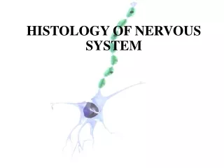
Morphology oftypical neuron • Neuronfunctional cell of the nervous tissue. • Cell body or perikaryon - contains the nucleus – regulates the functioning of the neuron. • Numerous dendrites and a single axon. • Contains Nissl bodies in the cytoplasm • Axon hillock- no Nissl bodies • Axon – cellular process (extension) – carries impulses away from the cell body. • Dendrites – cellular process (extension) – carries impulses toward the cell body
2 types of cell i) Neurons (nerve cells) ii) Supporting cells Functions of neurons specialized to receive stimuli and to conduct electrical impulses to other parts of the system. Arranged as an integrated communications network, with several neurons in a chain-like fashion involved in sending impulses from one part of the system to another.
Neuron Classification • Structural: • Multipolar – most common type in CNS. • Include all motor neurons and interneurons of brain and spinal cord. • Bipolar- not as common purely sensory. • Retina of eye, inner ear, olfactory epithelium in the upper region of nose. • Unipolar (formerly known as pseudounipolar) • Sensory neurons found in numerous craniosacral ganglia of the spinal cord.
Unipolar neuron Nucleus & nucleolus Cytoplasm fibrocytes Satellite cells Cytoplasm of neuron Myelinated axons
The supporting cells (neuroglia or glia): • Astrocytes • Oligodendrocytes • Microglial cells • Ependymal cells • Schwann cells • Satellite cells CNS PNS
Astrocytes • Largest, most numerous, versatile, and highly branched glial cells • They cling to neurons and cover capillaries • Functionally, they: • Support and brace neurons • Anchor neurons to their nutrient supplies • Guide migration of young neurons • Control the chemical environment
Microglia • Microglia – smallest, ovoid cells with spiny processes - phagocytic cells that migrate through the CNS and remove foreign and degenerated material
Ependymal Cells • Ependymal cells – squamous- to columnar-shaped cells • They line the central cavities of the brain and spinal column
Oligodendrocytes • Oligodendrocytes – branched cells that wrap CNS nerve fibers - Produce myelin in CNS
Schwann Cells and Satellite Cells • Schwann cells (neurolemmocytes) – form myelin sheaths around peripheral axons • Satellite cells surround neuron cell bodies with ganglia
Synapse • The region where the terminals come close to another cell and transmit the impulse • A junction that mediates information transfer from one neuron: • To another neuron • To an effector cell • Presynaptic neuron – conducts impulses toward the synapse • Postsynaptic neuron – transmits impulses away from the synapse
Myelin – formation & function • Whitish, fatty (protein-lipid), segmented sheath around most long axons • Its function: • Protection of the axon • Electrically insulating fibers from one another • Increasing the speed of nerve impulse transmission
2 types of neuroglia produce myelin • CNS= Oligodendrocyte • PNS= Schwann cells