Download
1 / 21
281 likes | 1.39k Views
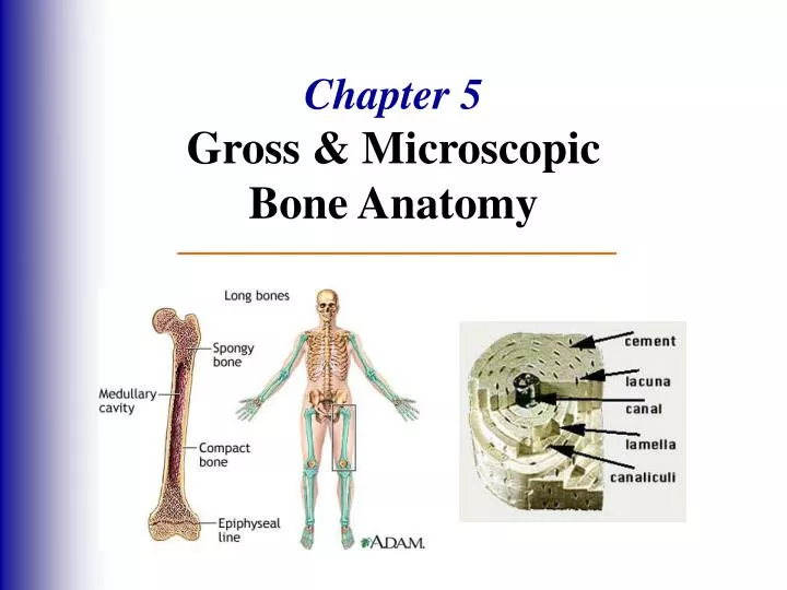
Chapter 5 Gross & Microscopic Bone Anatomy. The Skeletal System. Parts of the skeletal system Bones (skeleton) Joints Cartilages Ligaments Divided into 2 divisions Axial skeleton Appendicular skeleton. Functions of Bones. Support body Protect soft organs

E N D
The Skeletal System Parts of the skeletal system • Bones (skeleton) • Joints • Cartilages • Ligaments Divided into 2 divisions • Axial skeleton • Appendicular skeleton
Functions of Bones • Support body • Protect soft organs • Movement due to attached skeletal muscles • Storage of minerals and fats • Blood cell formation
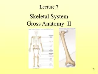
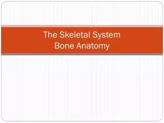
Bones of the Human Body The skeleton has 206 bones Two types of bone tissue • Compact bone - Homogeneous • Spongy bone - Small needle-like pieces of bone - Many open spaces
Paranasal Sinuses • Hollow parts of bones surrounding nasal cavity • Functions: • Lighten the skull • Resonate and amplify voice
The Hyoid Bone • Only bone that doesn’t articulate with another bone • Moveable base for tongue
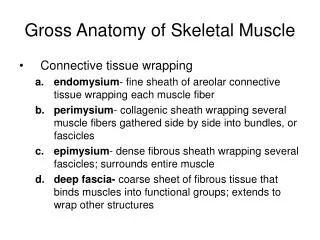
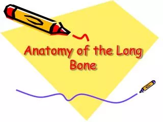
Gross Anatomy of a Long Bone Know 7 Structures 1. Diaphysis • Shaft • compact bone 2. Epiphysis • Ends of the bone • mostly spongy bone
3. Periosteum • Outside covering of diaphysis • Fibrous connective tissue membrane 4. Sharpey’s fibers • Secure periosteum to bone 5. Arteries • Supply bone cells with nutrients
6. Articular cartilage • Covers the ends • hyaline cartilage • Decreases friction 7. Medullary cavity • Cavity of shaft • Contains yellow marrow (mostly fat) in adults • Contains red marrow (for blood cell formation) in infants
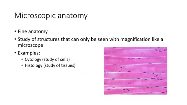
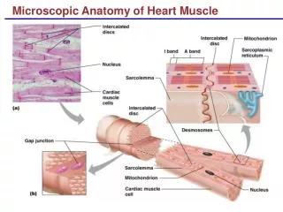
Microscopic Anatomy of Bone Know 6 parts: 1. Osteon (Haversian System) • A unit of bone 2. Central (Haversian) canal • Opening in the center • For blood vessels & nerves 3. Perforating (Volkmann’s) canals • perpendicular to central canal • For blood vessels & nerves
4. Lacunae • Cavities containing bone cells (osteocytes) • Arranged in rings 5. Lamellae • Rings around central canal 6. Canaliculi • Tiny canals • Radiate from central canal to lacunae • Transport system
Changes in the Growing Skeleton • In embryos, the skeleton is hyaline cartilage • During development, much of this cartilage is replaced by bone -Cartilage remains in isolated areas • Bridge of the nose • Parts of ribs • Joints
Bone Growth Epiphyseal (growth) plates - growth of long bone during childhood - New cartilage is continuously formed - Older cartilage ossified (broken down & replaced by bone)
Remodeled & lengthened - until growth stops - Bones change shape somewhat & grow in width
Types of Bone Cells 1. Osteocytes • Mature bone cells 2. Osteoblasts – b for build • Bone-forming cells 3. Osteoclasts – hard c for kill • Bone-destroying cells • Break down bone matrix for remodeling and release of calcium • Bone remodeling is a process by both osteoblasts and osteoclasts
Developmental Aspects • At birth, skull bones incomplete • Bones joined by fibrous membranes – fontanelles • Fontanelles replaced by bone within 2 years • Fetal skull is large compared to total body length
Bone Fractures • A break in a bone • Types 1. Closed (simple) fracture –does not penetrate skin 2. Open (compound) fracture –penetrates through skin • Bone fractures are treated by reduction and immobilization - Realignment of the bone
Repair of Bone Fractures 1. Hematoma (blood-filled swelling) is formed 2. Break is splinted by fibrocartilage to form a cartilaginouscallus 3. Fibrocartilage callus is replaced by a bony callus 4. Bony callus is remodeled to form a permanent patch