Download
1 / 19
490 likes | 985 Views
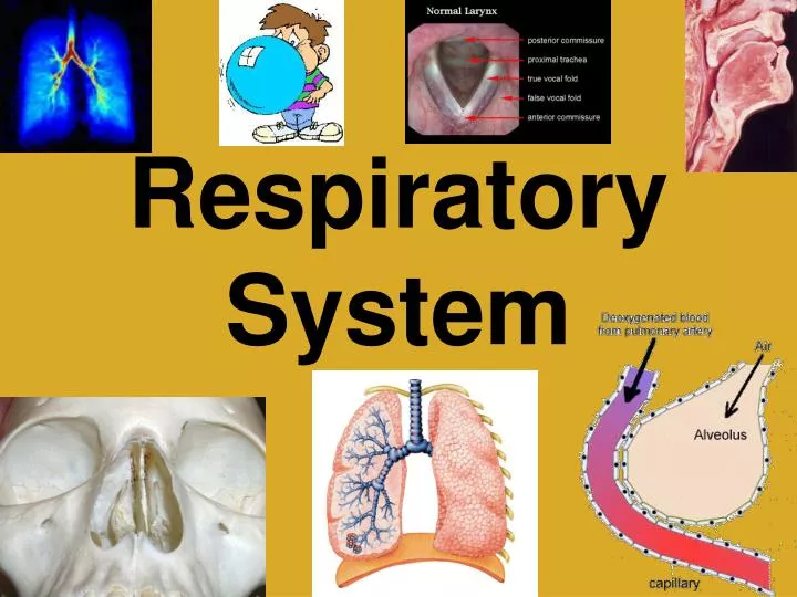
Respiratory System. I. Respiration. Process filters incoming air and transports it into the microscopic alveoli where gases are exchanged. Consists of the following: Ventilation Gas exchange between blood and lungs Gas transport in the bloodstream

E N D
I. Respiration • Process filters incoming air and transports it into the microscopic alveoli where gases are exchanged. • Consists of the following: • Ventilation • Gas exchange between blood and lungs • Gas transport in the bloodstream • Gas exchange between the blood and body cells • Cellular respiration
II. Organs of the Respiratory System • Divided into two groups: • Upper respiratory tract • Nose • Nasal cavity • Sinuses • Pharynx • Lower respiratory tract • Larynx • Trachea • Bronchial tree • Lungs
III. Upper Respiratory Tract • Nose- Filters air with coarse hairs inside the nostrils. • Nasal Cavity • Divided medially by the nasal septum. • Nasal Conchae • Divide the cavity into passageways (increase surface area) • Lined with mucous membrane to warm and filter incoming air.
III. Upper Respiratory Tract • Paranasal Sinuses • Air-filled spaces within the maxillary, frontal, ethmoid, and sphenoid bones of the skull. • Open to the nasal cavity • Lined with mucus membrane • Continuous with lining of the nasal cavity. • Reduce the weight of the skull • Serve as a resonant chamber to affect the quality of the voice. • Pharynx- Aids in producing sounds for speech.
IV. Lower Respiratory Tract • Larynx • Enlargement superior to the trachea and inferior to the pharynx. • Keeps particles from entering the trachea and also houses the vocal cords. • Vocal cords. • The upper pair is the false vocal cords. • The lower pair is the true vocal cords. • During swallowing, the false vocal cords and epiglottis close off the glottis.
IV. Lower Respiratory Tract • Trachea • Extends downward from larynx and splits into right and left bronchi. • Inner wall lined with ciliated mucous membrane (many goblet cells) to trap incoming particles. • The tracheal wall is supported by 20 incomplete cartilaginous rings.
IV. Lower Respiratory Tract • Bronchial Tree • Branched tubes leading from the trachea to the alveoli. • Begins with the two primary bronchi, each leading to a lung. • Primary Bronchi Bronchioles Alveolar Ducts Alveoli • Gas exchange between the blood and air occurs through the thin epithelial cells of the alveoli.
IV. Lower Respiratory Tract • Lungs • Spongy, cone-shaped • Enclosed by the diaphragm and thoracic cage. • The bronchi and large blood vessels enter each lung. • The right lung has three lobes, the left has two. • Lobes are composed of lobules
V. Breathing Mechanism • Ventilation (breathing)- composed of inspiration and expiration. • Inspiration • Diaphragm and intercostal muscles contract • Air pressure inside the lungs is decreased by increasing the size of the thoracic cavity. • Higher pressure air flows in from the outside.
V. Breathing Mechanism • Expiration • Diaphragm relaxes • Elastic recoil of lung and muscle tissues • Also from the surface tension within the alveoli. • Forced expiration is aided by thoracic and abdominal wall muscles.
VI. Respiratory Air Volumes and Capacities • The measurement of different air volumes is called spirometry. • Air that enters and leaves the lungs during one respiratory cycle is the tidal volume. • Maximum forced inspiration is the inspiratory reserve volume. • Maximum forced expiration is the expiratory reserve volume.
VI. Respiratory Air Volumes and Capacities • Vital capacity = tidal volume + inspiratory + expiratory reserve. • A residual volume always remains in the lungs. • Vital capacity + residual volume = total lung capacity. • Anatomic dead space is air remaining in the bronchial tree.
VII. Control of Breathing • Factors Affecting Breathing • Chemicals • Lung tissue stretching • Emotional state • Respiratory Center • Groups of neurons in the brain stem. • Central chemoreceptors • Sensitive to changes in the blood concentration of CO2 and [H+] • CO2 or [H+] concentrations = the breathing rate
VII. Control of Breathing • Peripheral chemoreceptors • In the carotid sinuses and aortic arch. • Sense changes in blood oxygen concentration. • Low O2 increases breathing rate and tidal volume increase. • Hyperventilation lowers the amount of carbon dioxide in the blood.
VII. Control of Breathing • Alveolar Gas Exchanges • Alveoli- Tiny sacs at the distal ends of the alveolar ducts. • Respiratory Membrane • Components • Epithelial cells of the alveolus • Endothelial cells of the capillary • The two fused basement membranes of these layers. • Gas exchange occurs across this respiratory membrane.
VIII. Gas Transport • Oxygen Transport • Over 98% of oxygen is carried in the blood bound to hemoglobin, producing oxyhemoglobin. • Oxyhemoglobin is unstable and gives up its oxygen in low O2 areas. • More oxygen is released as CO2 increases, as the blood becomes more acidic, and as blood temperature increases. • A deficiency of oxygen reaching the tissues is called hypoxia.
VIII. Gas Transport • Carbon Dioxide Transport • Bound to RBC as carbaminohemoglobin • Dissolved in plasma as bicarbonate ions. • The enzyme carbonic anhydrase is in RBCs • Enzyme combines CO2 with water forming carbonic acid
VIII. Gas Transport • Carbonic acid dissociates, releasing bicarbonate and hydrogen ions. • Most CO2 is transported as bicarbonate. • Carbaminohemoglobin releases its CO2 which diffuses out of the blood into the alveolar air.