Download
1 / 53
600 likes | 1.24k Views
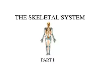
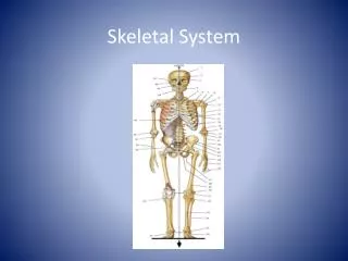
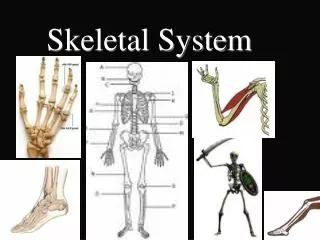
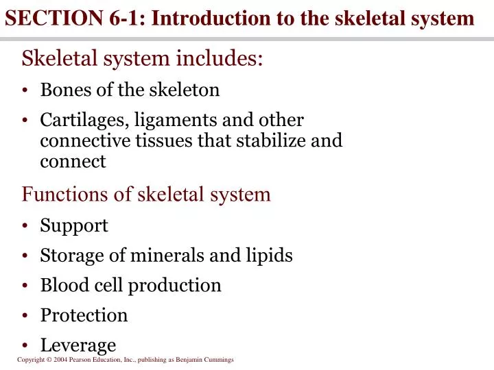
SECTION 6-1: Introduction to the skeletal system . Skeletal system includes: Bones of the skeleton Cartilages, ligaments and other connective tissues that stabilize and connect Functions of skeletal system Support Storage of minerals and lipids Blood cell production Protection Leverage.

E N D
SECTION 6-1:Introduction to the skeletal system Skeletal system includes: • Bones of the skeleton • Cartilages, ligaments and other connective tissues that stabilize and connect Functions of skeletal system • Support • Storage of minerals and lipids • Blood cell production • Protection • Leverage
SECTION 6-2:Classification of Bones • Long • Flat • Short • Irregular • Sesamoid • Sutural Bone shapes Bone Structure • Compact bone (dense) • Spongy bone (cancellous)
Figure 6.1 Classification of Bones by Shape Figure 6.1
A typical long bone includes • Diaphysis • Epiphyses • Metaphysis • Articular cartilage • Marrow cavity • Filled with red or yellow marrow • Articularcartlilage
Figure 6.2 Bone Structure Figure 6.2
SECTION 6-3: Bone Histology Osseous tissue • solid matrix- Crystals of hydroxyapatite Ca3(PO4)2+ Ca(OH)2 • Collagen- • Vascular-
SECTION 6-3: Bone Histology 4. Wrapped a. Endosteum b. Periosteum i. fibrous layer ii. Cellular layer(osteogenic)- iii. Sharpey’s Fibers
Cells in bone: • Osteocytes = mature bone cells -Maintain, repair bone 2. Osteoblasts= synthesize new matrix - become osteocyte 3. Osteoclasts=dissolve bone matrix -multicuncleated, differentiate from WBC 4.Osteoprogenitor =cells differentiate into osteoblasts, repair fractures
Figure 6.3 The Histology of Compact Bone Figure 6.3a
Figure 6.3 The Histology of Compact Bone Figure 6.3b, c
Compact bone and spongy bone • Basic unit of compact bone is an osteon • Osteocytes arranged around a central canal • Perforating canals extend between adjacent osteons • Spongy bone contains trabeculae
Figure 6.4 The Structure of Osseus Tissue Figure 6.4
Figure 6.5 The Distribution of Forces on a Long Bone Bones and stress • Compact bone located where stresses are limited in direction • Spongy bone located where stresses are weaker or multi-directional Figure 6.5
SECTION 6-4: Bone development and growth • Ossification = converting other tissue to bone 1. Intramembranous 2. Endochondral • Calcification = depositing calcium salts within tissues
deposition of Ca++ salts w/in a tissue Calcification
1. Intramembraneous Ossification • Bones form w/in a thin CT membrane • Used to form flat bones (cranium, mandible, clavicle)
forming bone: red cartilage: blue Intramembranous ossification • Begins with osteoblast differentiation • Dermal bones produced • Begins at ossification center
Steps of Intramembraneous Ossification in cranium • Mesenchymal cells form a thin CT membrane covering brain • Some mesenchymal cells differentiate into osteoblasts in the center of each membrane.(Occurs 6 weeks after fertilization) • Osteoblasts secrete bone @ these locations(called primary centers of ossification) • Ossification spreads in all directions
Steps of Intramembraneous Ossification in cranium • @ birth ossification is not complete, membrane remains as sutures and fontanels(soft spots) Fetal skulls at 13-34 weeks gestation; fontanels typically close at 6 months
Fontanelles • indentations of fibrous membrane between bones of fetal skull • Intramembranous ossification is not complete until 20-22 months • anterior • posterior • sphenoid • mastoid
Figure 6.7 Intramembranous Ossification Figure 6.7
Endochondral ossification • Cartilage model gradually replaced by bone at metaphysis • Increasing bone length • Timing of epiphyseal closure differs • Appositional growth increases bone diameter
Bone Growth 1. Appositional- increases diameter, while medullary cavity hollowed out -
Figure 6.10 Appositional Bone Growth Figure 6.10b
Bone Growth 2. Interstitial- occurs at epiphyseal plates a. Chondroblasts produce new cells that are pushed toward epiphysis b. At the diaphysis end of plate older cartilage is converted into bone http://www.personal.psu.edu/staff/m/b/mbt102/bisci4online/bone/bone_growth4.swf
Figure 6.9 Bone Growth at an Epiphyseal Cartilage Figure 6.9
SECTION 6-5: Dynamic Nature of Bone continually changing • Remodeling • Exercise • Hormone levels • Growth hormone and thyroxine increase bone mass • Calcitonin and PTH control blood calcium levels
Figure 6.12 A Chemical Analysis of Bone Figure 6.12
The skeleton is a calcium reserve • 99% body’s calcium in the skeleton • Calcium ion concentration maintained by bones GI tract and kidneys • Calcitonin and PTH regulate blood calcium levels • Calcitonin decreases blood calcium levels • PTH increases blood calcium levels
Effects on bone growth • Growth hormone- produced by pituitary gland; targets growth plate activity a. Excess causes giagantism
b. Low levels cause pituitary dwarfism • normally proportioned • treatment with growth hormone
Effects on bone growth 2. Testosterone- produced by the testes and adrenal glands in males and by the adrenal glands in females -Responsible for the growth “spurt’ at puberty -Overproduction in males causes closure of the epiphyseal line (Caution for anabolic steroid users)
Effects on bone growth • Estrogen- produced by the ovaries in females; -stimulates osteoblasts -promotes closing of the epiphyseal line -MENOPAUSE causes bone loss!!!
SECTION 6-7: Aging and the Skeletal System Effects of aging include 1. Osteopenia (inadequate ossification) begins between ages 30 and 40, • osteoblast activity declines, while osteoclast activity remains level, • Women lose 8% of bone mass per decade, while men lost only 3% in the same time period 2. Osteoporosis
Osteoporosis When the loss of bone mass compromises normal function a person is diagnosed with osteoporosis • When the loss of bone mass compromises normal function a person is diagnosed with osteoporosis.
Figure 6.16 The Effects of Osteoporosis Figure 6.16
Effects on bone growth 4. Parathyroid hormone- produced by the parathyroid glands in the neck -Stimulates the osteoclasts to Ca++ to be released from the bone to enter the blood -Ca++ necessary for muscles and nerves to function properly
Figure 6.13 Factors that Alter the Concentration of Calcium Ions in Body Fluids Figure 6.13a
Effects on bone growth 5. Calcitonin- produced by the thyroid gland - Inhibits osteoclasts so Ca++ remains in the bone , promotes ostepoblasts
Figure 6.13 Factors that Alter the Concentration of Calcium Ions in Body Fluids Figure 6.13b
Effects on bone growth 6. Vitamin D- some is produced in the skin ; Most is ingested in food -Necessary for the absorption of Ca++ from the intestine into the blood • -Osteomalacia (Rickets in children) • causing softened, weakened bones • Main symptom is pain when weight is put on the affected bone
Bone Fractures (Breaks) Bone fractures are classified by: Simple(closed) vs. compound(open)
Types of Bone Fractures • Linear • the fracture is parallel to the long axis of the bone • Transverse • the fracture is perpendicular to the long axis of the bone
Types of Bone Fractures • Greenstick • incomplete fracture where one side of the bone breaks and the other side bends • common in children
Types of Bone Fractures • Comminuted • bone fragments into three or more pieces • common in the elderly
Types of Bone Fractures • Spiral • ragged break when bone is excessively twisted • common sports injury
Types of Bone Fractures • Depressed • broken bone portion pressed inward • typical skull fracture
Types of Bone Fractures • Compression • bone is crushed; common in porous bones