Download
1 / 130
1.3k likes | 1.31k Views
Learn about the skeletal, muscular, and integumentary systems and their functions in the human body. Discover the structure and importance of bones in supporting the body and protecting internal organs.

E N D
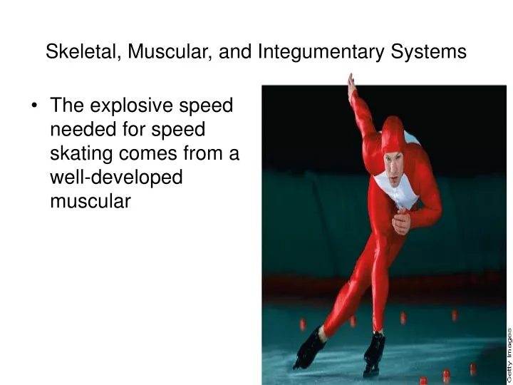
Skeletal, Muscular, and Integumentary Systems • The explosive speed needed for speed skating comes from a well-developed muscular
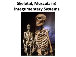
The Skeletal System • To retain their shapes, all organisms need some type of structural support • Single-celled organisms have a cytoskeleton that provides structural support • In multicellular animals, support is provided by some form of skeleton, including the external exoskeletons of arthropods and the internal endoskeletons of vertebrates • The human skeleton is composed of a type of connective tissue called bone • Bones and other connective tissues, such as cartilage and ligaments, form the skeletal system
The Skeletal System • Scientists can infer a lot about the behavior of extinct species by studying fossil bones and reconstructing skeletons • The human skeleton also contains important clues • The shape of your hip bones shows that you walk upright on two legs • The structure of the bones in your hands, especially your opposable thumbs, indicates that you have the ability to grasp objects • The size and shape of your skull is a clue that you have a well-developed brain
The Skeleton • The skeletal system has many important functions • The skeleton supports the body, protects internal organs, provides for movement, stores mineral reserves, and provides a site for blood cell formation • The bones that make up the skeletal system support and shape the body much like an internal wooden frame supports a house • Just as a house could not stand without its wooden frame, the human body would collapse without its bony skeleton • Bones protect the delicate internal organs of the body • For example, the skull forms a protective shell around the brain, and the ribs form a basketlike cage that protects the heart and lungs
The Skeleton • Bones provide a system of levers on which muscles act to produce movement • Levers are rigid rods that can be moved about a fixed point • In addition, bones contain reserves of minerals, mainly calcium salts, that are important to many body processes • Finally, bones are the site of blood cell formation • Blood cells are produced in the soft marrow tissue that fills the internal cavities in some bones
SKELETON • Human body contains 206 bones • Human skeleton is an internal structure referred to as a Endoskeleton • Bones: • Support the muscles and organs • Give shape and structure to the body • Store calcium and phosphorous which are important minerals used by the body in certain metabolic processes • Internal portions of certain bones manufacture blood cells • Protect delicate internal organs • Cranium (skull): protects the brain • Ribs: curved bones that form a cage to protect the heart and lungs
The Skeleton • There are 206 bones in the adult human skeleton • These bones can be divided into two parts: • Axial skeleton: supports the central axis of the body • Consists of the skull, the vertebral column, and the rib cage • Appendicular skeleton: • The bones of the arms and legs, along with the bones of the pelvis and shoulder area
SKELETON STRUCTURE • Composed of two parts: • Axial skeleton: consisting of about 80 bones, including the spine, ribs, sacrum, sternum, and cranium • Appendicular skeleton: contains 126 bones, including the bones of the arms, legs, pelvis, and shoulders
The Human Skeleton • The skeleton supports the body • The human skeleton is divided into two parts: • Axial skeleton • Appendicular skeleton
Structure of Bones • It is easy to think of bones as nonliving • After all, most of the mass of bone is mineral salts—mainly calcium and phosphorus • However, bones are living tissue • Bones are a solid network of living cells and protein fibers that are surrounded by deposits of calcium salts
Structure of Bones • The figure at right shows the structure of a typical bone • The bone is surrounded by a tough layer of connective tissue called the periosteum • Blood vessels that pass through the periosteum carry oxygen and nutrients to the bone • Beneath the periosteum is a thick layer of compact bone • Although compact bone is dense, it is far from being solid • Running through compact bone is a network of tubes called Haversian canals that contain blood vessels and nerves
Structure of Bones • Bones are a solid network of living cells and protein fibers that are supported by deposits of calcium salts • A typical long bone such as the femur contains spongy bone and compact bone • Within compact bone are Haversian canals,which contain blood vessels
BONE STRUCTURE • Bones are made up of both organic and inorganic material • Internal structure of long bones (arms/legs) • Periosteum: outer membrane of the bone which contains a network of blood vessels that supplies living bone cells with oxygen and nutrients and carries away carbon dioxide • Compact bone: • Under the periosteum • Hard material • Composed of rings of mineral crystals and protein fibers • Osteocytes: • Living bone cells interspersed throughout the mineral rings of the Compact Bone • Haversian Canal: • Channel in the center of each mineral ring in the Compact Bone containing nerves and blood vessels • Spongy bone: • Connective tissue interior to the Compact Bone and at end of long bones • Lacy structure adds strength to the bone without adding weight • Bone marrow: • Red: • Consist of blood vessels, fibers, cells • Manufactures erythrocytes and white blood cells • Found in spongy bone /ends of long bones/ribs/vertebrate/sternum/pelvis • Yellow • Consist mostly of fat cells • Serves as energy reserve • Fill shafts of the long bones
Structure of Bones • A less dense tissue known as spongy bone is found inside the outer layer of compact bone • It is found in the ends of long bones and in the middle part of short, flat bones • Despite its name, spongy bone is not soft and spongy; it is actually quite strong • Near the ends of bones where force is applied, spongy bone isorganized into structures that resemble the supporting girders in a bridge • This latticework structure of spongy bone helps to add strength to bone without adding mass
Structure of Bones • Osteocytes, which are mature bone cells, are embedded in the bone matrix • Two other kinds of bone cells—osteoclasts and osteoblasts line the Haversian canals and the surfaces of compact and spongy bone • Osteoclasts break down bone • Osteoblasts produce bone • Although we stop growing in our late teens, our bones are continuously remodeled through the activity of osteoclasts and osteoblasts
Structure of Bones • Within bones are cavities that contain a soft tissue called bone marrow • There are two types of bone marrow: • Yellow: • Made up primarily of fat cells • Red: • Produces red blood cells, some kinds of white blood cells, and cell fragments called platelets
Development of Bones • The skeleton of an embryo is composed almost entirely of a type of connective tissue called cartilage • The cells that make up cartilage are scattered in a network of protein fibersincluding both tough collagen and flexible elastin • Unlike bone, cartilage does not contain blood vessels • Cartilage cells must rely on the diffusion of nutrients from the tiny blood vessels in surrounding tissues • Because cartilage is dense and fibrous, it can support weight, despite its extreme flexibility
Development of Bones • Cartilage is replaced by bone during the process of bone formation called ossification • Ossification begins to take place up to seven months before birth • Bone tissue forms as osteoblasts secrete mineral deposits that replace the cartilage in developing bones • When the osteoblasts become surrounded by bone tissue, they mature into osteocytes
Development of Bones • Many long bones, including those of the arms and legs, have growth plates at either end • The growth of cartilage at these plates causes the bones to lengthen • Gradually, this new growth of cartilage is replaced by bone tissue, and the bones become larger and stronger • During late adolescence or early adulthood, the cartilage in the growth plates is replaced by bone, the bones become completely ossified, and the person “stops growing”
Development of Bones • In adults, cartilage is found in those parts of the body that are flexible, such as the tip of the nose and the external ears • Cartilage also is found where the ribs are attached to the sternum, which allows the rib cage to move during breathing
BONE DEVELOPMENT • Ossification: process by which bones develop • Two types: • Long bones first develop as cartilage that is later replaced by bone • Cartilage: tough, flexible connective tissue • Once osteocytes develop in the cartilage, they releaseminerals that lodge in the spaces between cartilage cells eventually replacing the cartilage • Some cartilage is never replaced: disc between vertebrae, joints, end of nose, external ear, trachea • Makes area flexible • Other bones develop directly from embryonic connective tissue without forming cartilage first • Clavicle, some parts of the skull
Types of Joints • A place where one bone attaches to another bone is called a joint • Joints permit bones to move without damaging each other • Some joints, such as those of the shoulder, allow extensive movement • Others, like the joints of the fully developed skull, allow no movement at all • Depending on its type of movement, a joint is classified as immovable, slightly movable, or freely movable
JOINTS • Location where two bones meet • Ligaments: tough bands of connective tissue that holds the bones of a joint in place • Stretch as the bones move • Three kinds: • Fixed: skull • Semimovable(bend and twist): vertebral column, ribs • Movable: • Hinge (back and forth movement): elbow • Ball and socket (circular motion): hip, shoulder • Pivot (side to side / up and down): cervical vertebrae • Angular (twisting): wrists, ankles • Gliding (slide against each other): small bones of the hand and feet
Immovable Joints • Immovable joints, often called fixed joints, allow no movement • The bones at an immovable joint are interlocked and held together by connective tissue, or they are fused • The places where the bones in the skull meet are examples of immovable joints
Slightly Movable Joints • Slightly movable joints permit a small amount of restricted movement • Unlike the bones of immovable joints, the bones of slightly movable joints are separated from each other • The joints between the two bones of the lower leg and the joints between adjacent vertebrae are examples of slightly movable joints
Freely Movable Joints • Freely movable joints permit movement in one or more directions • Freely movable joints are grouped according to the shapes of the surfaces of the adjacent bones
Freely Movable Joints • Ball-and-socket joints permit movement in many directions • They allow the widest range of movement of any joint • Hinge joints permit back-and-forth motion, like the opening and closing of a door • Pivot joints allow one bone to rotate around another • Saddle joints permit one bone to slide in two directions
Types of Freely Movable Joints • Freely movable joints are classified by the type of movement they permit • The joints illustrated are in the shoulder, knee, elbow, and hand
FUNCTIONING OF JOINTS • Joints are subject to a great deal of pressure and stress • Connective tissue near the joints secretes synovial fluidwhich cushions the bones thus preventing the bones from wearing away • Fluid filled sac called the bursais found in the knee and shoulder joints adding an additional cushion between the bones
Structure of Joints • In freely movable joints, cartilage covers the surfaces where two bones come together • This protects the bones as they move against each other • The joints are also surrounded by a fibrous joint capsule that helps hold the bones together while still allowing them to move
Structure of Joints • The joint capsule consists of two layers • One layer forms strips of tough connective tissue called ligaments • Ligaments, which hold bones together in a joint, are attached to the membranes that surround bones • Cells in the other layer of the joint capsule produce a substance called synovial fluid, which forms a thin film on the cartilage that covers the bony surfaces that form the joint • This lubricating film enables the surfaces of the joint to slide over each other smoothly
Structure of Joints • In some freely movable joints, such as the knee in the figure, small sacs of synovial fluid called bursae (singular: bursa) form • A bursa reduces the friction between the bones of a joint and also acts as a tiny shock absorber.
Knee Joint • The knee joint is protected by cartilage and bursae • The ligaments hold the bones composing the knee joint—femur, patella, tibia, and fibula—together
Skeletal System Disorders • Bones and joints can be damaged, just like any other tissue • Excessive strain on a joint may produce inflammation, a response in which excess fluid causes swelling, pain, heat, and redness • Inflammation of a bursa is called bursitis • A more serious disorder is arthritis, which involves inflammation of the joint itself • Arthritis affects approximately 10 percent of the world's population
JOINT INJURY • Bursitis: inflammation of the bursa • Sprain: torn ligament • Arthritis: • Synovial membranes become inflamed and grow thicker • Fibrous tissues in the joints may become ossified, immobilizing the joint and fusing the bones together
Skeletal System Disorders • Because bone is living tissue, calcium is moved between it and the rest of the body to maintain homeostasis of this important mineral • In older people, especially women, loss of calcium can lead to a weakening of the bones, a condition known as osteoporosis • Osteoporosis can cause many serious fractures • Sound nutrition, including plenty of calcium in the diet, and weight-bearing exercise are among the best ways to prevent this serious problem
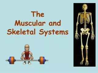
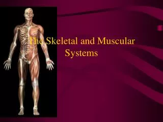
The Muscular System • Despite the fantasies of Hollywood horror films, a skeleton cannot move by itself • Movement is the function of the muscular system • More than 40 percent of the mass of the average human body is muscle • The muscular system includes the large muscles displayed by some athletes • It also includes thousands of tiny muscles throughout the body that help to regulate blood pressure, move food through the digestive system, and power every movement of the body—from the blink of an eye to the hint of a smile
Types of Muscle Tissue • Muscle tissue is found everywhere in the body—not only just beneath the skin but also deep within the body • There are three different types of muscle tissue: skeletal, smooth, and cardiac • Each type of muscle is specialized for a specific function in the body
MUSCLE • Contractile organ consisting of many cells • Three types: • Skeletal • Cardiac • Smooth
Muscle Tissue • There are three types of muscle tissue: skeletal, smooth, and cardiac • Skeletal muscle cells have striations, or stripes, and many nuclei • Smooth muscle cells are spindle-shaped and have one nucleus and no striations • Cardiac muscle cells have striations and usually only one nucleus