Download
1 / 64
650 likes | 1.14k Views
Neoplasms of Lung and Pleura. William K. Funkhouser, M.D. Ph.D. x 3-1069 Bill_Funkhouser@med.unc.edu. Neoplasms of Lung and Pleura. Primary Neoplasms of Lung Primary Neoplasms of Pleura Metastatic Neoplasms to Lung and/or Pleura. Neoplasms of Lung and Pleura: Classification by Lineage.

E N D
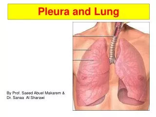
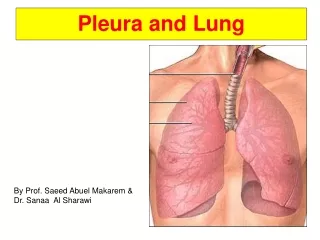
Neoplasms of Lung and Pleura William K. Funkhouser, M.D. Ph.D. • x 3-1069 • Bill_Funkhouser@med.unc.edu
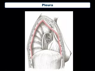
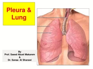
Neoplasms of Lung and Pleura • Primary Neoplasms of Lung • Primary Neoplasms of Pleura • Metastatic Neoplasms to Lung and/or Pleura
Neoplasms of Lung and Pleura: Classification by Lineage • Epithelial – most common • Melanocytic • Stromal • Mesothelial
Benign Lung Neoplasms • Hamartoma • Squamous papillomatosis • Pleomorphic adenoma (ENT)
Hamartoma • Clin: Adolescence adulthood • None in newborns - not congenital • Rad: Solitary nodule +/- popcorn calcification • Peripheral > central • Path: Gross: solitary, lobulated, cartilagenous Micro: normal tissues in excess/disarray
Solitary Pulmonary Nodule Hamartoma
Bivalved Hamartoma
Cartilage in excess and disarray Hamartoma
Malignant epithelial neoplasms(Carcinomas) • Squamous cell carcinoma • Adenocarcinoma • Large cell undifferentiated carcinoma • Small cell undifferentiated carcinoma
Lung Carcinomas:Epidemiology • Estimated Incidence (2003): 172,000 (US) • Estimated Mortality (2003): 157,000 (US) • >85% of lung carcinoma deaths (and 30% of all cancer deaths) occur in cigarette smokers • Risk = f(# cigarettes smoked), 15-30X in heavy smokers, 50-60X in asbestos workers who smoke • Risk decreases with cessation of cigarette smoking: baseline after 15 years
USA Tobacco Use • 25% of US adults smoke cigarettes • M=F • US adults consume 2,400 cigs/person/year • 36% of US high school students smoke • est. 1.8 million new smokers/year (65% < 18 yo)
Tobacco: Morbidity and Mortality • Premature ASVD: major risk factor • Emphysema: Linear with exposure: 7%/10 years • Chronic bronchitis • Carcinomas of pharynx, larynx, lung, esophagus, bladder, kidney • Fetal tobacco syndrome
Tobacco: Chemistry • 80% air, 20% gases and particulates • Gases: CO, CO2, formaldehyde, acrolein, methanol, phenol, anthracenes, pyrenes • Nicotine: 1% of smoke 85% absorbed in lung equivalent to 1 mg IV
Tobacco: Chemistry • Particulates: • resin cores in 0.5 M diameter water droplets • est. 109 particles/ml • 50% deposited in and cleared by cilia • remainder: phagocytosis, lymphatic transport • Overall: 4,000 chemical compounds, of which 43 are considered carcinogenic
Squamous cell carcinoma • Clin: Smokers (98%) 20-30% of common carcinomas May secrete PTH-like compound • Rad: central > > peripheral • Path: Bronchi > Larynx > Trachea +/- Desmosomes (intercellular bridges) +/- Keratin production, e.g. keratin pearls
Squamous cell carcinoma in situ Respiratory mucosa
Desmosomes Keratin Invasive Squamous Carcinoma
Normal lymph node lymphocytes Mets in subcapsular sinuses Metastatic squamous cell carcinoma to lymph node
Adenocarcinoma • Clin: 30-40% of common carcinomas Most common carcinoma in non-smokers, but 80% of adenoCAs occur in smokers • Rad: peripheral > central • Path: +/- glands +/- mucin Bronchiolo-alveolar carcinoma subset
Primary Pleural effusion Adenocarcinoma
Gland formation Adenocarcinoma
Mucin production (red on PASd stain) Adenocarcinoma
Bronchioloalveolar carcinoma (BAC) • Clin: Rising incidence (presently 20-25%) Not associated with cigarette smoking • Rad: Peripheral, can be multifocal and bilateral • Path: Lepidic (butterfly-like) growth pattern Mucinous or non-mucinous Unifocal or multifocal Distinction of multifocal primary from mets
Large cell undifferentiated carcinoma • Clin: 10% of common carcinomas • Rad: non-specific • Path: H&E: Undifferentiated EM: ? adenocarcinomas cDNA microarrays: distinct disease
Non-Small Cell Lung Carcinomas: Prognostic variables • Definitely: Stage, performance status, weight loss • Possibly gender, ploidy, k-ras mutation, p53 protein accumulation • Not age, histology
Small cell carcinoma • Clin: Smokers 20 % of common carcinomas Ectopic ACTH, ADH, Eaton-Lambert, carcinoid s. Commonly high stage at presentation Responsive to chemo/RT, but low 5 yr survival • Rad: Central in >90% Frequent metastases to LNs and distant sites • Path: Malignant cytology No nucleoli High mitotic activity and necrosis
Response to therapy At diagnosis Small cell undifferentiated carcinoma
Necrotic carcinoma Viable carcinoma Small cell undifferentiated carcinoma
Normal lymphocytes Metastatic small cell carcinoma
Small Cell Lung Carcinoma: Prognostic variables • Definitely: Stage, performance status • Probably: Gender, age, # of metastatic sites
Neoplasms of Lung & Pleura: Classification by Lineage • Epithelial • Melanocytic • Stromal • Mesothelial • Metastases
Mesothelioma • Clin: Associated with asbestos exposure • Rad: Diffuse pleural involvement May have associated effusion • Path: Malignant Deeply invasive growth pattern Epithelial, spindle cell, or biphasic Immuno: Keratin (+) EM: long microvilli
Visible C-P Angle Loss of C-P Angle = Pleural effusion or mass Mesothelioma: PA Chest
Normal thickness pleura Thickened pleura Mesothelioma: CT
Epithelioid cytology of this mesothelioma mimics adenocarcinoma
Adenocarcinoma Mesothelioma N. Weidner