Download
1 / 48
480 likes | 675 Views
Lecture 19 Reproductive Systems. Somatic cells (human) have _______ pairs chromosomes Diploid vs Haploid Division by _______________ Gamete production: Division by _____________ Number of division cycles __________ Number of hypothetical resulting cells________

E N D
Lecture 19 Reproductive Systems
Somatic cells (human) have _______ pairs chromosomes • Diploid vs Haploid • Division by _______________ • Gamete production: • Division by _____________ • Number of division cycles __________ • Number of hypothetical resulting cells________ • Ploidy level in daughter cells_____________ • Opportunities for new genetic combinations from:
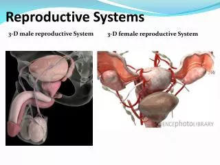
Male Reproductive System • Scrotum • Spermatic cord • Testes • Histology • Accessory organs • Penis
Scrotum • Two cavities • Midline seam = ________________ • Visceral membrane: • Tunica vaginalis • Muscles • Cremaster muscle • Dartos muscle
Spermatic cord • Passage from peritoneal cavity to scrotal chambers • Extends through ________________ • Contains: • ____________ • Testicular artery • Pampiniform plexus ____________ • Function:
Testes • Connective tissue covering ______________ • Lobules formed by partitions • Seminiferous tubules • Interstitial cells produce ___________ • Temperature regulation – how? • Muscles: • counter-current cooling mechanism: • Blood testes barrier • Reason • Maintained by:
Sperm production • Sperm production: mitosis of spermatogonium spermatocyte • Meiosis: spermatocyte spermatids • Physical maturation: sperm • Blood-testes barrier • FSH & Testosterone • Nurse cells (= Sertoli or sustenacular cells) • Promote and regulate process • inhibin • Sperm anatomy • Flagella • Acrosome • head
Male Reproductive Tract • Epididymis • Regions • Function • Ductus deferens • Ampulla • Ejaculatory duct
Accessory Organs - • Seminal glands (vesicles) • Prostate gland • Bulbo-urethral glands
Penis • Regions: • ____________________ • _____________________ • _____________________ • Prepuce = foreskin – often removed • Erectile tissues • _____________________ • _____________________
Sperm Morphology • Adapted for reaching & penetrating a secondary oocyte • Head contains DNA & acrosome (hyaluronidase and proteinase enzymes) • Midpiece contains mitochondria to form ATP • Tail is flagellum used for locomotion
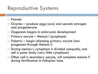
Female Reproductive System • Designed for release of one gamete/month • Includes measures for preparation on uterine wall to receive fertilized egg • Organs include: • Ovaries produce 2nd oocytes & hormones • Uterine tubes transport fertilized ova • Uterus where fetal development occurs • Vagina & external genitalia constitute the vulva • Mammary glands produce milk
What is released at ovulation? When does meiosis start? When is meiosis completed?
Internal Supporting ligaments: • Broad ligament • Mesovarium • Round ligament • Ovarian ligament • Suspensory ligament
Ovary Structure • Outer layer: ___________________ • Site of gamete production • Gametes formed within ______________ • Inner layer: medulla • Covered by: • Germinal epithelium • Tunica albuginea
Oogenesis • Occurs within a _____________________ • Oogonia – 2n stem cell • Mitosis prior to birth • primary oocytes • Located within _______________________ • Follicle development/oocyte division stimulated by __________________________ • Ovulation releases ___________________ • Surrounded by cell layer = _________________________________ • Corpus luteum n corpus albicans
Uterine Tubes • ~ 5 inches • Parts: • Infundibulum • Fimbriae • Ampulla • Isthmus • Uterine part • Walls - ________________ muscle • Peristaltic contractions • Lined with _________________________
Uterus • Fundus • Body • Isthmus • Cervis • Internal and external os • Histology: • Myometrium • Endometrium • perimetrium
Change in Histology – Uterine Cycle • Menses • Proliferative stage • Secretory phase
Hysterectomy • Surgical removal of the uterus • Indications for surgery • endometriosis, ovarian cysts, excessive bleeding, cancer of cervix, uterus or ovaries • Complete hysterectomy removes cervix • Radical hysterectomy removes uterus, tubes, ovaries, part of vagina, pelvic lymph nodes and supporting ligaments
Hormonal Control of Ovarian and Menstrual Cycles From Anterior Pituitary • FSH stimulates development of follicle in ovary • Luteinizing hormone – triggers ovulation From Ovaries (follicle cells/corpus luteum) • Estrogen – preparation of uterine lining for implantation • Progesterone – maintenance of endometrium
Vulva • Mons pubis -- fatty pad over the pubic symphysis • Labia majora & minora -- folds of skin encircling vestibule where find urethral and vaginal openings • Clitoris -- small mass of erectile tissue • Bulb of vestibule -- masses of erectile tissue just deep to the labia on either side of the vaginal orifice
Mammary Glands • Modified sweat glands that produce milk (lactation) • amount of adipose determines size of breast • milk-secreting glands open by lactiferous ducts at the nipple • areola is pigmented area around nipple • suspensory ligaments suspend breast from deep fascia of pectoral muscles (aging & Cooper’s droop)
Terminology of Development • Gestation period • time span from fertilization to birth (38 weeks) • Prenatal period (before birth) • embryological development • developing human for first 2 months after fertilization is known as an embryo • all principal adult organs are present • fetal development • from 9 weeks until birth is known as a fetus • by end of 3rd month, placenta is functioning • Neonatal period is first 42 days after birth • obstetrics is branch of medicine that deals with the neonatal period, pregnancy and labor
Fertilization • Occurs as sperm penetrate follicular layers • Corona radiata • Zona pellucida • Fuses with haploid nucleus of oocyte • Various mechanisms prevent penetration of a second sperm to egg • Fertilized egg is termed a _____________
Development of the Blastocyst • A blastocyst is a hollow ball of cells that enters the uterine cavity by day 5 • outer covering of cellscalled the trophoblast • inner cell mass • fluid-filled cavity calledthe blastocele • Trophoblast & part of innercell mass will develop into fetal portion of placenta • Most of inner cell mass will become embryo
Implantation • Attachment of blastocyst to endometrium • occurs 6 days after fertilization • implants with inner cell mass in contact with the endometrium • Trophoblast develops 2 distinct layers • syncytiotrophoblast secretes enzymes that digest the endometrial cells • cytotrophoblast is distinct layer of cells that defines the original shape of the embryo • Trophoblast secretes human chorionic gonadotropin (hCG) that helps the corpus luteum maintain the uterine lining
Ectopic Pregnancy • Blastocyst implants outside uterus • Possible sites include:\ • Cervix • Ovary • Fallopian tubes • Membranes surrounding reproductive organs • Cause: often impaired passage in fallopian tubes • Twice as common in smokers because nicotine paralyzes the cilia • Possibly fatal to mother
Development from Blastocyst • Inner cell mass gives rise to embryo • Folding of inner wall (Gastrulation) results in organization of: • Ectoderm • Mesoderm • Endoderm
A multicellular organism develops through a series of embryonic stages adult • Development continues to death • Growth – cell division/expansion • Differentiation specialized cell types • Morphogenesis – creation of overall form through pattern formation
Embryonic Induction • a change in developmental fate of an embryonic tissue through exposure to gene product (morphogen) from adjacent tissue
Morphogen: • Chemical signals that diffuse from inducing tissues to nearby tissues • Gradient formed – cells at various distances respond differently • Switch on blocks of genes direct formation of body plan
Formation of Embryonic Membranes • Yolk sac • site of early blood formation • gives rise to gonadal stem cells (spermatogonia & oogonia) • Amnion • surrounds embryo with fluid: shock absorber, regulates body temperature & prevents adhesions • fluid is filtrate of mother’s blood + fetal urine • examine a sample of it for embryonic cells (amniocentesis) • Chorion • becomes the embryonic contribution to the placenta • derived from trophoblast & mesoderm lining it • gives rise to human chorionic gonadotropin (hCG) • Allantois • outpocketing off yolk sac that becomes umbilical cord
Placenta & Umbilical Cord • Placenta forms during 3rd month • chorion of embryo & stratum functionalis layer of uterus • Chorionic villi extend into maternal blood filled intervillous spaces --- maternal & fetal blood vessels do not join & blood does not mix • diffusion of O2, nutrients, wastes • stores nutrients & produces hormones • barrier to microorganisms, except some viruses • AIDS, measles, chickenpox, poliomyelitis, encephalitis • not a barrier to drugs such as alcohol • Placenta detaches from the uterus (afterbirth)
Fetal Circulation • Corionic villi – provide exchange surface at placenta • Mother and child blood never mix – but nutrients and toxins cross • Umbilical cord – blood route to and from placenta where exchange takes place
Path of Fetal Blood • Oxygen/nutrient rich blood returns to fetus in umbilical vein • Since no oxygen exchange take place in fetal lungs Oxygen rich blood shunts to left atrium via oval opening between atria • Oval opening closed by flap at birth • Atrial blood shunts to aorta (from right ventricle to aorta)
Summary of Development • 1st trimester • 2nd trimester • 3rd trimester