Download
1 / 25
260 likes | 381 Views
Learn about the cutaneous nerves, muscles, deep fascia, veins, and arteries in the posterior aspect of the leg. Understand the divisions, functions, and innervation of various muscle groups for a detailed study.

E N D
Cutaneous Nerves • Saphenous n. (from femoral n.) • Posterior femoral cutaneous n.(from sacral plexus) • Lateral cutaneous n. of leg (from common fibular (peroneal) n.) • Sural n.(from tibial n & common fibular (peroneal) n.)
Superficial veins • Smallsaphenousvein • piercestheroof of thepopliteal fossa anddrainsintothepoplitealvein
Deep fascia of leg(Fascia cruris) • interosseous membrane and the “crural intermuscular septa” divide the leg into 3 crural compartments: 1-Anterior comp. 2-Lateral comp. 3-Posterior comp.: contains 2 group of muscles: • superficial • deep
A L P
Flexor Retinaculum • broad band of deep fascia which passes -from medial malleolus -to calcaneus
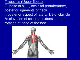
Muscles Superficial layer • Gastrocnemius m. • Soleus m. • Plantaris m. Deep layer • Popliteus m. • Flexor digitorum longus m. • Flexor hallucis longus m. • Tibialis posterior m. insert to calcaneus with a common tendon “tendo calcaneus” (achille’s tendon) pass deep to the “flexor retinaculum”
SUPERFICIAL LAYERGastrocnemius m. • Mostsuperficial of themuscles in theposteriorcruralcompartment • O: 2 heads,lat& med condyles of femur, theycometogether at theinferiormargin of thepopliteal fossa • I: Calcaneous, via “tendocalcaneus” (aschillestendon) • N: Tibial n. • F: Flexion (plantarflexion) of foot & leg
Soleus m. -Located deep to gastrocnemius • O: Soleal line, upper part of fibula • I: calcaneus via “tendo calcaneus” • N: Tibial nerve • F: Flexion of foot Gastrocnemius+soleus mm: Triceps surae m.
Plantaris m. ---Small muscle; variable in size and extend, it may be absent. • O: Lat supracondylar area of the femur • I: Medial part of tendo calcaneus • N: Tibial n. • F: assistsflexion of leg, plantar flexion of the foot
DEEP LAYER:Popliteus m. ---Thin, triangular muscle • O: Lat condyle of femur & arcuate popliteal lig. • I: Post surface of tibia • N: Tibial nerve • F: Flexion & medial rotation of leg
Flexor digitorumlongusm. • O: Post surface of tibia below soleal line • I:Bases of distal phalanges of 2-5 • N.:Tibial nerve • F: Flexion of toes 2-5 & foot, inversion of foot, helps to maintain the medial longitudinal arch of foot
Flexor hallucislongus m. --Thelong, powerfulandlargestmuscle of deeplayer • O: Lower part of post surface of fibula, interosseosmemb. • I: Base of distal phalanx of hallux (bigtoe of foot) • N.: Tibialnerve • F: Flexion of hallux & foot, alsoimportant in holding theleg in the normal position of foot.
Tibialisposterior m. • Thedeepest m in theposteriorcruralcompartment • O: Lateral part of post surface of tibia below soleal line, post surface of fibula, interosseous membrane • I: Sustentaculumtali, tuberosity of navicular b, talus, cuneiform bb, cuboid b, 2-4metatarsal bb • N.: Tibial n. • F: Plantar flexes andinverts foot cross
Posterior tibial artery • it begins at thedistalborder of thepopliteusmuscle • it is larger terminal branch of thepoplitealartery • it is accompaniedbythetibialnerve • Branches: • Fibular ( peroneal) a • Circumflex fibular a • Medial malleolar a • Calcanealbrs • Nutrient a of tibia • Lat & med plantar aa
Fibular (peroneal) artery • Muscular branches • Nutrient a. to fibula • Perforating br. • Communicating br. • Lateral malleolar brr. • Calcaneal brr.