Download
1 / 27
300 likes | 801 Views
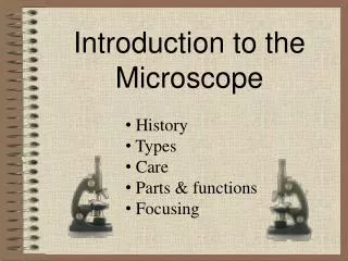
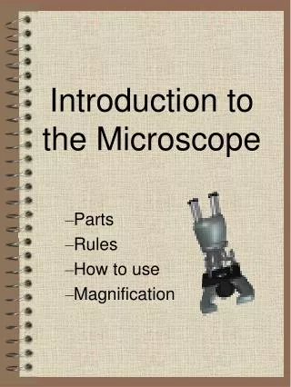
Introduction to the Light Microscope. VCE Biology Induction. Eye piece (Ocular). Revolving nose piece(Turret). Objective lenses (x40,x10,x4). Specimen holder. Coarse adjustment knob. Stage. Iris Diaphragm . Fine adjustment knob. Condenser. lamp.

E N D
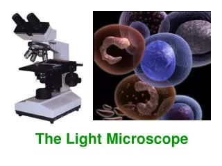
Introduction to the Light Microscope VCE Biology Induction
Eye piece (Ocular) Revolving nose piece(Turret) Objective lenses (x40,x10,x4) Specimen holder Coarse adjustment knob Stage Iris Diaphragm Fine adjustment knob Condenser lamp Specimen holder Y axis adjustment knob Main switch Light intensity adjustment knob Specimen holder X axis adjustment knob
Adjust iris diaphragm to match the magnification of the objective lens in place
Units of measurement • Light microscope: micrometers are generally used • Micrometer=micron • Symbol for micrometer is µm 1cm = 10mm 1mm =1000 µm 1cm = 10000µm
Introducing the mini- grid 1000 µm 1mm 1000 µm 1mm 1cm 10mm 10mm 10000 µm 1cm 10000 µm
A closer look at the mini grid 0.5 mm= 500 µm 100 µm 1000 µm
Field of view Entire Field of view Diameter of field of view Low power Diameter of FOV ~1800 µm High power Diameter of FOV ~450 µm
1000µm x 40 objective lens (x400 mag) HIGH POWER Field of view
Estimating Size and Scale • Ensure that you know the magnification • For the magnification used recall the diameter of the field of view • Count how many cell fit across the field of view and record this figure • Count how many cells fit up/down
Estimating size of cell Magnification x 100 Diameter of FOV~ 1800 µm 6 cell fit across Width of one cell 18006= 300 µm So width of one cell is ~ 300 µm
1 2 3 4 5 6 7 8 38 µm Scale calculation Diameter of FOV~ 1800 µm InventedCell: Unstained x 100 Recall:18006= 300 µm So width of one cell is ~ 300 µm cytoplasm Diagram width 8cm 300 µm 8 = 37.5 So 1 cm = 38 µm nucleus Cell membrane
x 100 Invented Cell: Unstained Final Drawing (Diameter of FOV~ 1800 µm) cytoplasm nucleus Cell membrane 38 µm Scale (Diagram width 8cm) 300 µm8 = 37.5 So 1 cm = 38 µm Size18006= 300 µm So width of one cell is ~ 300 µm
Onion Cells viewed under high power Recall: Diameter of F.O.V ~ 450µm How many cell fit across FOV ? ~2 1/3 cells across Length of one cell 450 2.3=195.65 µm How many cell fit down FOV? ~6 cell fit down One onion cell is approx 196µm in length and 75µm in width Width of one cell 4506= 75µm
nucleus 1 2 3 4 5 6 7 8 Scale calculation Onion Cells: Stained with Methylene Blue Mag x 400 Diameter of FOV~ 450 µm Recall:4502.3=196 µm So length of one cell is ~ 196 µm Diagram length of cell 7.5cm : 196µm 7.5= 26.1 So 1 cm =26 µm 26 µm
nucleus 26 µm What should final diagram look like? ( x 400) Onion Epidermis Cells Stain:Methylene Blue ACTUAL LENGTH OF CELL Diameter of FOV~ 450 µm SCALE Diagram length of cell 7.5cm 450 2.3=195.7 µm So length of one cell is ~ 196 µm 196 µm7.5= 26 µm So 1 cm =26 µm
BIOLOGICAL DRAWING • Drawing materials • Positioning • Size • Accuracy • Technique • Labelling drawing • Title/size/scale conventions
Transverse section Cell sections Longitudinal section