Download
1 / 43
430 likes | 588 Views
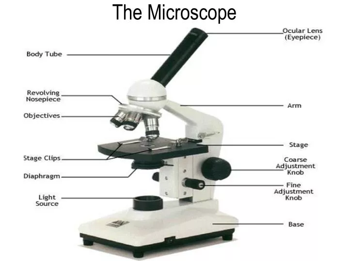
The Microscope. or light source. The Microscope. Scientist use microscopes to reveal details that otherwise might be difficult or impossible to see Biologist use them to study organisms and their parts. Simple Microscope.

E N D
The Microscope or light source
The Microscope • Scientist use microscopes to reveal details that otherwise might be difficult or impossible to see • Biologist use them to study organisms and their parts
Simple Microscope • The simple microscope is a single lens microscope is used to produce an enlarged image.
Compound Microscope • Compound Microscope- shines light through specimen and has at least two lenses to magnify an image.
The Development of Light Microscopes Section 7.1 Summary – pages 171-174 Compound light microscopes can magnify objects up to 2,000 times.
The Microscope • Magnification- the increase of an object’s apparent size • Power of magnification is the degree of enlargement (how many times will the object be multiplied?) • Power of magnification = ocular (eyepiece) lens X magnification of objective lens
The Microscope • Resolution- the ability of the microscope to deliver a clear image **the higher the magnification the lower the resolution
Development of Electron Microscopes • The electron microscope was invented in the 1940s. • This microscope uses a beam of electrons to magnify structures..
Transmission Electron Microscope (TEM) • Transmission Electron Microscope (TEM)- • a beam of electrons must pass through a very thinly sliced specimen. • Has great resolution of internal structures • Can magnify objects up to 500,000 times. • The transmission electron microscope (TEM) allows scientists to study the structures contained within a cell.
Scanning Electron Microscopes (SEM) • Scanning Electron Microscope- • passes beams of electrons over (scans the surface of) the specimen’s surface and provides a 3D image • Can be magnified up to 100,000 times.
Father of Microscopy • Anton Van Leeuwenhoek (1632-1723) • Leeuwenhoek is known to have made over 500 "microscopes," of which fewer than ten have survived to the present day. These typically magnified between 20 to 30 times. • He discovered blood cells, and was the first to see living sperm cells of animals. He discovered microscopic animals.
What is a cell? • A cell is the smallest unit of life that can carry on all the functions of life.
Who first discovered cells? • Robert Hooke first discovered cells in 1665 • He was an English scientist that looked at a cork oak tree with an early light microscope; they reminded him of the rooms or “cells” where monks lived.
Who first observed living cells? • Anton van Leeuwenhoek in 1673 • He viewed pond water organisms and called them “Wee beasties”. We now call them protists.
What are the three parts of the cell theory? 1. All living things are made of cells 2. Cells are the basic units of structure & function in all organisms. 3. Cells come only from the reproduction of existing cells.
What shapes do cells come in? • Cells come in a variety of shapes and their shapes usually reflect their function. • Examples: (we will do the chart together)
How big or small are cells? -Cells are measure in micrometers(µm) -Most cells are only 10-50µm in diameter. -The smallest cell is bacteria -The largest cell is ostrich egg -The longest cell is nerve cell in a giraffe’s leg
How big can cells get? • The size of a cell is limited by the relationship of the cell’s outer surface area to it’s volume • (Surface area to volume ratio)
Surface Area • A two-dimensional measurement of the size of the surface of an object
Volume • The amount of three dimensional space occupied by an object.
SA to Volume Ratio • As cells grow, their volume increases much more than their surface area! • The limited surface area would not allow materials to enter or leave the cell quickly enough to meet the cell’s needs.
Surface Area to Volume • What would happen if you fried the whole potato instead of making French fries?
What are the three main parts? 1. Cell Membrane- semi or selectively permeable. It controls which substances pass in and out of the cell. Made up of a phospholipid bilayer. 2. Nucleus- contains the hereditary information (DNA) and directs most of the cell’s activities. 3. Cytoplasm- the region between the cell’s nucleus and the cell membrane, which contains the organelles and the cytosol (jelly-like substance)
What are the two types of cells? • Eukaryote - cells that have a nucleus and membrane-bound organelles. • Examples: plant and animal cells • Prokaryote cells that do not have a nucleus and membrane-bound organelles. • Examples:bacteria
How do plant and animal cells differ? PLANT • Cell wall • Central vacuole • Chloroplast • No lysosomes ANIMAL • No cell wall • No central vacuole • No chloroplast • Lysosomes
What is the difference between unicellular and multicellular organisms? • Unicellular- one cell can carry out all the functions of life. Example: bacteria, paramecium, amoeba • Multicellular – organisms composed of many cells that are specialized to carry out specific jobs (Cell Specialization) • Therefore the cells depend upon other cells for survival (Cell Interdependence)
What are the levels of structural organization in multicellular organisms? • Atom and molecule • Macromolecules • Organelles 4. Cell 5. Tissue 6. Organ 7. Organ System 8. Organism
More pictures Tissue Organ
Organ Organ Systems
Cells Product • All cells must generate protein (monomer: amino acid) • Cells make protein through a process called protein synthesis.
Cells Product • Transcription: the process of making a copy of DNA into mRNA. This occurs in the nucleus • Translation: the process of translating mRNA to tRNA into an amino acid chain.