Download
1 / 14
150 likes | 278 Views
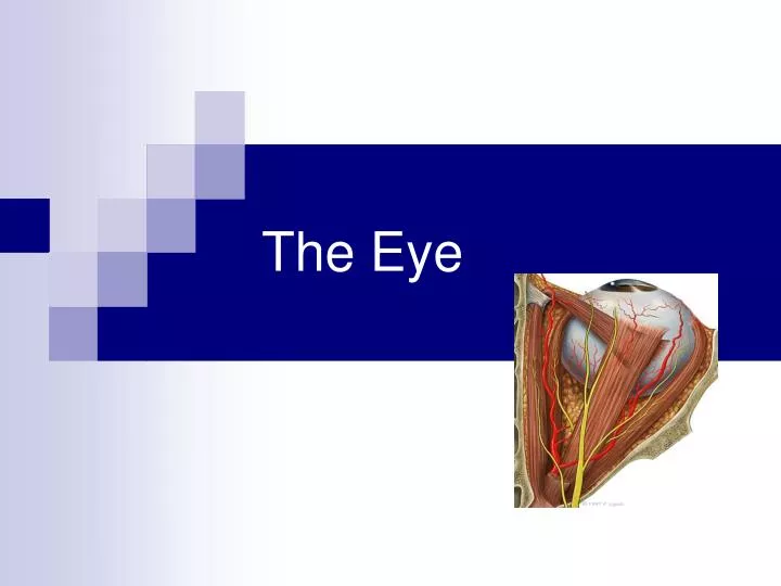
The Eye. Structure of the Eyeball. Slightly irregular hollow sphere with anterior and posterior poles The wall is composed of three tunics- fibrous, vascular and sensory The internal cavity is filled with fluids called humors.

E N D
Structure of the Eyeball • Slightly irregular hollow sphere with anterior and posterior poles • The wall is composed of three tunics- fibrous, vascular and sensory • The internal cavity is filled with fluids called humors. • The lens separates the internal cavity into anterior and posterior segments
Fibrous Tunic • Forms the outermost coat of the eye: • Opaque sclera (posterior) • Clear cornea (anterior) • Sclera protects the eye • Cornea lets light enter the eye
Vascular Tunic (Uvea): Choroid Region • Has three regions: • Choroid • Cilliary body • Iris • Choroid Region: • A dark brown membrane that forms the posterior portion of the uvea • Supplied blood to all eye tunics
Vascular Tunic: Cilliary Body • Thickened ring of tissue surrounding the lens • Composed of smooth muscle bundles • Anchors the suspensory ligament that holds the lens in place
Vascular Tunic: Iris • The colored part of the eye • Pupil- central opening of the iris • Regulates amount of light entering the eye during: • 1. close vision and bright light- pupil contracts • 2. distant vision and dim light- pupil dilates • 3. changes in emotional state- pupils dilate when the subject matter is appealing or requires problem solving skills
Sensory Tunic: Retina • A delicate two-layered membrane • Pigmented layer- the outer layer absorbs light and prevents scattering • Neural Layer, which contains: • Photoreceptors that transduce light energy • Bipolar cells and ganglion cells • Amacrine and horizontal cells
The Retina: Ganglion cells and the Optic Disc • Ganglion cell axons: • Run along the inner surface of the retina • Leave the eye as the optic nerve • The optic disc: • Is the site where the optic nerve leaves the eye • Lacks photoreceptors (blind spot)
The Retina: Photoreceptors • Rods: • Respond to dim light • Are used for peripheral vision • Cones: • Respond to bright light • Have high-acuity color vision • Are found in the macula lutea • Are concentrated in the fovea centralis
Blood Supply to the Retina • The neural retina receives its blood supply from two sources • The outer third receives its blood from the choroid • The inner two-thirds are served by the central artery and vein • Small vessels radiate out from the optic disc and can be seen with an ophthalmoscope.