Download
1 / 39
390 likes | 421 Views
Dive into the impact of mechanical stress on bone healing, common fracture types, diagnostic techniques, and treatment options for optimal recovery.

E N D
Response to Mechanical Stress • Wolff’s law – a bone grows or remodels in response to the forces or demands placed upon it • Observations supporting Wolff’s law include • Long bones are thickest midway along the shaft (where bending stress is greatest) • Curved bones are thickest where they are most likely to buckle
Response to Mechanical Stress • Trabeculae form along lines of stress • Large, bony projections occur where heavy, active muscles attach
Statistics • Fractures of extremities most common • More common in men up to 45 years of age • More common in women over 45 years of age • Before 75 years wrist fractures (Colles’) most common • After 75 years hip fractures most common
Fractures A fracture is any break in a bone. • Fracture repair involves formation of a clot called a fracture hematoma, organization of the fracture hematoma into granulation tissue called a procallus (subsequently transformed into afibrocartilaginous [soft] callus), conversion of the fibrocartilaginous callus into the spongy bone of a bony (hard) callus, and, finally, remodeling of the callus to nearly original form.
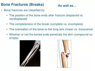
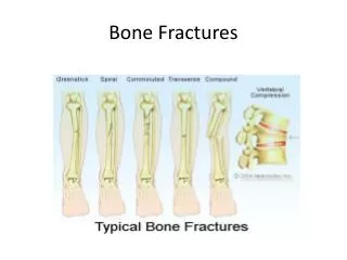
Bone Fractures (Breaks) • Bone fractures are classified by: • The position of the bone ends after fracture • The completeness of the break • The orientation of the bone to the long axis • Whether or not the bones ends penetrate the skin
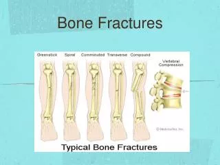
Common Types of Fractures • Magnitude and direction of force are determining factors in type of fracture. • Closed • – Bone fragments do not pierce skin • Open/compound • – Bone fragments pierce skin • Displaced or undisplaced
Common Types of Fractures • Comminuted – bone fragments into three or more pieces; common in the elderly • Spiral – ragged break when bone is excessively twisted; common sports injury • Depressed – broken bone portion pressed inward; typical skull fracture • Compression – bone is crushed; common in porous bones
Common Types of Fractures • Epiphyseal – epiphysis separates from diaphysis along epiphyseal line; occurs where cartilage cells are dying • Greenstick – incomplete fracture where one side of the bone breaks and the other side bends; common in children
Common Types of Fractures • Named for shape or position of fracture line • Common types of fracture • Pott’s -- distal fibular fracture • Colles’s -- distal radial fracture • stress fracture -- microscopic fissures from repeated strenuous activities
Transverse fracture • Usually caused by directly applied force to fracture site
Spiral (Oblique) • Caused by violence transmitted through limb from a distance (twisting movements)
Greenstick • Occurs in children: bones soft and bend without fracturing completely
Compression (Crush) fractures • Fracture in cancellous bone: result of compression (osteoporosis)
Burst fracture • Occurs in short bones, e.g. vertebra from strong direct pressure such as impaction of disc.
Avulsion fracture • Caused by traction, bony fragment usually torn off by a tendon or ligament. • What muscle group attaches to this bony prominence and what nerve also runs in close proximity? • Forearm flexors (common flexor origin) ulnar nerve
Fracture dislocation/subluxation • Fracture involves a joint: results in malalignment of joint surfaces.
Impacted fracture • Bone fragments are impacted into each other.
Comminuated fracture • Two or more bone pieces - high energy trauma
Comminuated fractures can require serious hardware to repair.
Stress Fracture • Abnormal stress on normal bone (fatigue fracture) or normal stress on abnormal bone (insufficiency fracture).
Functions of the X-ray • Localizes fracture and number of fragments • Indicates degree of displacement • Evidence of pre-existing disease in bone • Foreign bodies or air in tissues • May show other fractures • MRI, CT or ultrasound to reveal soft tissue damage
Healing is faster in bone than in cartilage due to lack of blood vessels in cartilage Healing of bone is still slow process due to vessel damage Clinical treatment closed reduction = restore pieces to normal position by manipulation open reduction = realignment during surgery Repair/Healing of Bone
How to Handle Fractures • Reduction • Open reduction – Allows very accurate reduction – Risk of infection – Usually when internal fixation is needed • Manipulation – Usually with anesthesia • Traction – Fractures or dislocation requiring slow therapy
Fracture Fixation • 4-12 weeks • External fixation • Internal fixation • Intermedually nails, compression plates • Frame fixation
External fixation • Used for fractures that are too unstable for a cast. You can shower and use the hand gently with the external fixator in place.
Frame fixation • Allows correction of deformities by moving the pins in relation to the frame.
Stages in the Healing of a Bone Fracture • Hematoma formation • Torn blood vessels hemorrhage • A mass of clotted blood (hematoma) forms at the fracture site • Site becomes swollen, painful, and inflamed • 3-4 hours Hematoma Hematoma formation 1
Stages in the Healing of a Bone Fracture • The fibrocartilaginous callus forms when: • Osteoblasts and fibroblasts migrate to the fracture and begin reconstructing the bone • Fibroblasts secrete collagen fibers that connect broken bone ends • Osteoblasts begin forming spongy bone • Osteoblasts furthest from capillaries secrete an externally bulging cartilaginous matrix that later calcifies
Stages in the Healing of a Bone Fracture • Fibrocartilaginous callus forms • Granulation tissue (soft callus) forms a few days after the fracture • Capillaries grow into the tissue and phagocytic cells begin cleaning debris External callus New blood vessels Internal callus (fibrous tissue and cartilage) Spongy bone trabeculae Fibrocartilaginous callus formation 2
Stages in the Healing of a Bone Fracture • Bony callus formation • New bone trabeculae appear in the fibrocartilaginous callus • Fibrocartilaginous callus converts into a bony (hard) callus • Bone callus begins 3-4 weeks after injury, and continues until firm union is formed 2-3 months later Bony callus of spongy bone Bony callus formation 3
Stages in the Healing of a Bone Fracture • Bone remodeling • Excess material on the bone shaft exterior and in the medullary canal is removed • Compact bone is laid down to reconstruct shaft walls Healing fracture Bone remodeling 4