Download
1 / 25
250 likes | 280 Views
Explore the pathophysiology, clinical manifestations, diagnosis, and imaging modalities of ARVD, a myocardial disorder primarily affecting the right ventricle with genetic implications and potential life-threatening arrhythmias.

E N D
Arrhythmogenic Right Ventricular Dysplasia Georgia Giakoumis Spear, M.D. April 10, 2007
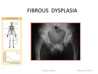
What is ARVD? • Myocardial disorder of primarily the RV • Characterized histologically by gradual replacement of myocytes by adipose and fibrous tissue
Epidemiology • Uncertain cause and prevalence • Idiopathic cardiomyopathy • Some report prevalence as 0.4% (Van der Wall EE et al.) or 1 in 5000 (Gemayel et al.) • Familial (30%) • Autosomal dominant inheritance with variable penetrance and incomplete expression • Recently, several genes have been implicated
Etiology • Many theories have been described • These include: • Apoptosis leading to progressive myocardial muscle loss followed by fibrofatty replacement • This results in electrical vulnerability of the RV which may lead to life threatening arrythmias • CHD-abnormal development of RV leading to dysplasia • Metabolic disorder affects the RV and fatty and fibrous replacement occurs • Healing process in context of myocarditis
Pathologic features of ARVD • Two variants: fatty and fibrofatty • Fatty • Replacement of myocardium without thinning of ventricular wall • Involves only the RV • Fibrofatty • Thinning of RV • May involve LV as well • Specifically, the septum, LV free wall with a predilection for the posteroseptal and posterolateral areas.
Anatomic findings • Mild to severe global dilitation of the ventricle • Ventricular aneurysms at the site of the “Triangle of Dysplasia” can be considered pathognomonic for ARVD • TRIANGLE OF DYSPLASIA • RV subtricuspid areas • Apex • infundibulum • Segmental hypokinesia
Clinical Picture • Predominantly young adults • M:F ratio is 2.7:1 • May result in sudden death • 20% of sudden deaths in <35yo • 22% of sudden deaths in young athletes
Clinical Picture • 80% of cases are diagnosed <40 yo • Patient presentation • Syncope • VT • Cardiac arrest • Adult patients with CHF
Clinical Picture • Spectrum of disease • Asymptomatic form: ventricular ectopic beats • Biventricular heart failure • +/- arrythmias • (ie. Ventricular arrythmias with LBBB (originates from RV) • High incidence of inducible supraventricular arrythmias • Possible sudden death • Temporal progression
EKG findings • Regular sinus rhythm • QRS>110msec in V1 • Epsilon wave beyond the QRS in V1 (30%) • Inversion of T waves in precordial leads V1-V3 (50%)
Manifestations of ARVD • Electrocardiographic repolarization and depolarization changes • Structural abnormalities ranging from subtle wall aneurysms within the “triangle of dysplasia” • Biventricular regional or global dysfunction • Localized or widespread fibrofatty infiltration of the RV myocardium
Diagnosis • Major and minor criteria which include: • Genetics • Electrocardiographic findings • Pathophysiologic phenomena • Histopathologic factors • Imaging, especially MR should be used as an important additional criteria (Kayser et al. Radiographics 2002; 22: 639-650) • Patients must have 2 major, 1 major and 2 minor or 4 minor to fulfill the appropriate criteria for ARVD
Van der Wall EE et al. MRI Findings in ARVD
DDX • Idiopathic dilated cardiomyopathy • Dilated CM vs ARVD • Generalized CM manifests as progressive decline in LV fxn • ARVD progressive decline in RV fxn • Uhl anomaly (paper-thin RV due to near complete absence of myocardial muscle fibers • Uhl anomaly vs ARVD. • Uhl anomaly has no gender predilection or familial occurrence • Usually presents in infancy with CHF
Imaging modalities which aid in the evaluation of the RV • Conventional angiography • Echocardiography • Radionuclide angiography • Ultrafast CT • MR—allows the clearest visualization of the heart
Imaging Evaluation • RV angiography is the standard of reference for diagnosis • Discerns abnormalities such as akinetic or dyskinetic bulging in infundibular, apical and subtricuspid regions • Echo • Used to exclude other anatomic abnormalities • However, these traditional methods lack sensitivity and specificity for detecting structural and functional abnormalities of the RV myocardium
MR • 3D evaluation of ventricular anatomy and volumes • Excellent spatial resolution • Unlimited FOV
MR Findings • T1- high signal intensity of fat in the RV myocardium • Fibrofatty replacement leading to diffuse thinning of the RV myocardium (major criteria) • RV and RVOT aneurysms (major criteria) • Dilation of the RV and RVOT (major criteria when severe; minor criteria when mild) *These findings should be considered a major criteria in the diagnosis of ARVD
More MR findings • Can also assess both systolic and diastolic function • RV diastolic dysfunction is an early marker of disease even with LV fxn is preserved • Regional contraction abnormalities (minor) • Global systolic dysfunction (major) • Global diastolic dysfunction (minor)
Therapy • Options include: • Antiarrythmic medication • Sotalol, Amiodarone or combination therapy • Goal to prevent recurrent ventricular tachycardia • Catheter ablation • Using RF or DC energy • Reserved for patients who are unresponsive or intolerant to the drugs • Patients may have new ventricular arrythmias • Implantable AICD • Surgery • Total disconnection of the RV
References: • Gemayel, C. et al. “Arrythmogenic Right Ventricular Cardiomyopathy.” Journal of the Americal College of Cardiology. 38 (2001)1773-1781 • Marcus et. Al. “Arrhymogenic right ventricular dysplasia/cardiomyopathy:a review.” Pacing Clinical Electrophysiology. 18 (1995) 1298-314 • Auffermann et al. “Arrhythmogenic Right Ventricular disease: MR Imaging vs Angiography.” AJR. 161 (1993) 549-555 • Kayser et al. “Diagnosis of Arrhythmogenic Right Ventricular Dysplasia: A Review.” Radiographics. 22 (2002) 639-650. • Van der Wall et al. “Arrythmogenic Right Ventricular Dysplasia: MRI Findings.” Herz Cardiovascular Disease. 25 (2000) 356-364.