Download
1 / 21
260 likes | 1.29k Views
Tympanoplasty, Mastoidectomy, Facial Nerve Decompression. Hau Sin Wong Grand Rounds 10/27/04. Tympanoplasty. Definition: operation involving tympanic membrane and evaluation of middle ear Whereas myringoplasty is an operation of the tympanic membrane

E N D
Tympanoplasty, Mastoidectomy, Facial Nerve Decompression Hau Sin Wong Grand Rounds 10/27/04
Tympanoplasty • Definition: operation involving tympanic membrane and evaluation of middle ear • Whereas myringoplasty is an operation of the tympanic membrane • Tympanoplasty can be accompanied with or without mastoidectomy
Types of Tympanoplasty • Type I: restoration of normal ME with intact ossicles • Type II: Ossicular chain partially destroyed but preserved ad continuity restored
Type III: TM lays on stapes suprastructure • Type IV: Round window protection with a small middle ear mobile footplate left exposed • Type V: Closed middle ear with round window protection; fenestra in the horizontal semicircular canal coveredby a skin graft.
Middle Ear Anatomy • Ossicles: Malleus, Incus, Stapes • Ligaments: sup malleolar ligament, stapedial tendon • Cochlearform process, pyramidal process
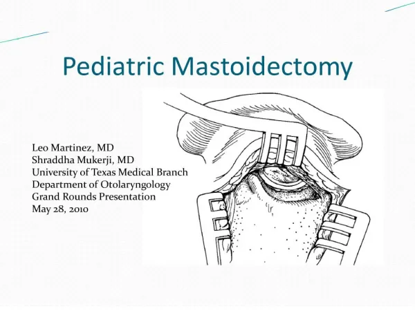
Technique • Evaluate TM and ME if perforation present • Create vascular strip • Incise postauricular incision for mastoidectomy • Mastoidectomy- identify lateral landmarks for drilling. Anterior boundary-post EAC, superior boundary- root of the zygoma, linea temporalis, inferior boundary- mastoid tip. Drill medially and saucerize, delineating Tegmen superiorly, thinning post EAC anteriorly, Sigmoid sinus posteriorly, Horizontal SCC medially • Identify vertical segment of facial nerve between HSCC and digastric ridge • Open facial recess: boundaries are fossa incudus, facial nerve, chorda tympani • Visualization of incus, malleus in antrum, incus and stapes in facial recess.
Tympanomeatal flap • EAC skin and TM elevated anteriorly
TM grafts: • 1. Cartilage 2. fool’s fascia (loose areolar fascia over temporalis fascia) 3. temporalis fascia • Medial or lateral graft placement
Anatomy 5 segments • Meatal- brainstem to IAC • Labyrinthine- fundus of IAC to facial hiatus (includes fallopian canal-narrowest segment of facial canal) • Tympanic- geniculate gangilion to pyraminal eminence • Mastoid-pyramidal eminence to stylomastoid foramen • Extratemporal- stylomastoid foramen to muscles of facial expression ( temporal, zygomatic, buccal, mandibular, cervical)
Anatomy • Arterial Supply: • Intracranial segment supplied by labyrinthine artery off the AICA • Tympanic segment supplied by superficial petrosal artery off the middle meningeal artery • Mastoid segment supplied by the stylomastoid artery off the external carotid artery
Facial Nerve Decompression • Eggshell the facial canal (vertical segment) • Open Facial recess and extended the facial recess using a barber pole method • Follow facial nerve to the tympanic segment • Eggshell covering overlying facial nerved removed, exposing the sheath • Sheath incised and facial nerve decompressed.