Download
1 / 32
330 likes | 364 Views
Learn about non-displaced hip fractures, challenging to diagnose on X-rays. Discover ultrasound features, importance of early detection, and imaging techniques. Screening ultrasound could prevent missed fractures.

E N D
Ultrasound diagnosis of non displaced hip fractures Dr Richard Beese Bsc (Hons) MRCP FRCR St Georges Hospital, London
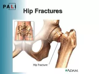
Non displaced fractures • Examples : Hip , scaphoid , rib sternum , some facial fractures, toddlers tibia fracture and greater tuberosity humeral fracture • All fractures can be non displaced and difficult to see on X-ray
Objectives • What are non displaced hip fractures and why are they difficult to diagnose ? • Why is it important to diagnose non displaced hip fractures ? • How we diagnose non displaced hip fractures. • The ultrasound features of fractures and imaging technique.
What are non displaced hip fractures ? Impacted fractures of the neck often in a subcapital position
Impacted fractures of the neck often in a subcapital position
Why are non displaced hip fractures difficult to diagnose ? • Not visible on x-ray, because of preservation of shentons line, no fracture line visible , hip trabeculae intact. • Patients can weight bare, straight leg raise and demonstrate normal axial compression.
70 year old lady fall onto her right side and admitted with severe right hip pain, failed to mobilise well on the ward , repeat pelvic xray 3 days later.
3 day interval repeat AP pelvis demonstrating a displaced sub capital fracture on the right.
75 year old lady fall onto left side. AP pelvis no fracture CT no fracture seen
Why is important to diagnose non displaced hip fractures early? No one wants to miss a fracture and send patients home with a broken hip. Increase immobility and thus morbidity Change of operation from a simple screw fixation to a hemiarthropasty
Post traumatic Painful Hip Sonography as a screening test for Occult hip fractures. Ori Safran MD et al Orthopaedic and Radiology Hadassah Hebrew University Medical Centre Jerusalem Israel Journal ultrasound med 2009;28:1447-1452 30 patients ultrasound of both hips compared with MRI. 100% sensitive 65% specific. Concluded, a negative ultrasound can exclude intra articular trauma.
50 year old man RTA left hip pain
Is this new ? • Elbow effusions and lipoheamathrosis of the Knee are used to diagnose intra atricular fractures on plain x-ray. • Ultrasound is demonstrating ‘ an anterior fat pad sign of the hip’.
Possible pathway in the presence of trauma No bone injury Refer for further Imaging
Conclusion Screening Ultrasound of the hip in trauma ; Has a short learning curve to become competent. Widely available in A/E and quick to perform at the bedside. Easy to interrupt, comparison both sides. US screening may prevent missing hip fractures in future
WHO; 25% of the worlds population have local access to x-rays • Ultrasound is being used to differentiate between • soft tissue and bone injury in remote austere locations. • e.g. Australia and Canada • Only 1/3 of patients in Darenth Valley Hospital fracture clinic have a fracture – Huge waste of time and money with immobilisation of soft tissuie injuries. • Ultrasound screening could used to improve this.