Download
1 / 60
600 likes | 748 Views
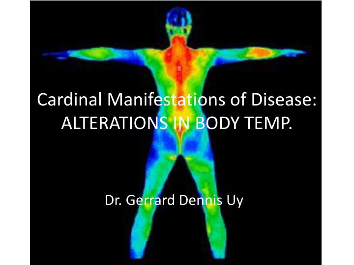
Cardinal Manifestations of Disease: ALTERATIONS IN BODY TEMP. Dr. Gerrard Dennis Uy. Fever and Hyperthermia. hypothalamic thermoregulatory center – regulates body temperature (core BT 37 C) A.M. temperature of >37.2°C (>98.9°F) or a P.M. temperature of >37.7°C (>99.9°F) would define a fever

E N D
Cardinal Manifestations of Disease: ALTERATIONS IN BODY TEMP. Dr. Gerrard Dennis Uy
Fever and Hyperthermia • hypothalamic thermoregulatory center – regulates body temperature (core BT 37 C) • A.M. temperature of >37.2°C (>98.9°F) or a P.M. temperature of >37.7°C (>99.9°F) would define a fever • Rectal temperatures are generally 0.4°C (0.7°F) higher than oral readings
Fever • Elevation of body temperature that exceeds the normal daily variation • occurs in conjunction with an increase in the hypothalamic set point • Vasoconstriction in hands and feet shunts blood to the internal orgrans • shivering = heat conversion and production • until the temperature of the blood bathing the hypothalamic neurons matches the new thermostat setting
Fever • Pathogenesis • Pyrogen – any substance that causes fever • Exogenous (microbial products, microbial toxins, or whole microorganisms) • E.g. LPS endotoxin – gram negative bacteria • Endogenous a.k.aPyrogenic cytokines (IL-1, IL-6, tumor necrosis factor (TNF), ciliaryneurotropic factor (CNTF), and interferon (IFN) ) • Elevation of Hypothalamic Set point • levels of prostaglandin E2 (PGE2) are elevated in hypothalamic tissue and the third cerebral ventricle
Hyperthermia • uncontrolled increase in body temperature that exceeds the body's ability to lose heat • setting of the hypothalamic thermoregulatory center is unchanged • does not involve pyrogenic molecules • Exogenous heat exposure and endogenous heat production
Hyperthermia • Can be rapidly fatal • Does not respond to antipyretics
The Decision to Treat Fever • Most fevers are associated with self limiting infections • There is no significant clinical evidence that antipyretics delay the resolution of viral or bacterial infections nor there is evidence that fever facilitates recovery from infection • Fever can provide important diagnostic clues in different disease conditions
The Decision to Treat Fever • The objective in treating fever is to reduce the elevated set point and to facilitate heat loss • Reducing fever also reduces systemic symptoms • Treatment of fever in some patients is highly recommended (cadiac, cerebrovascular, and pulmonary diseases)
Mechanism of antipyretics • Acts by reducing the level of PGE2 • Common antipyretics • Acetaminophen • Aspirin • NSAIDS • glucocorticoids
Approach to the patient with fever and rash • A thorough history if often needed • Includes: • Immune status • Medications taken within the previous month • Travel history • Immunzation status • Exposure to domestic pets • Recent exposure to ill individuals • The history should also include onset and direction and rate of spread
Types of lesions • Macules - flat lesions defined by an area of changed color • Papules - raised, solid lesions <5 mm in diameter • plaques ->5 mm in diameter with a flat, plateau-like surface • nodules ->5 mm in diameter with a rounded configuration • Wheals (urticaria, hives) - papules or plaques that are pale pink and may appear annular (ringlike) as they enlarge • Vesicles (<5 mm) and bullae (>5 mm) are circumscribed, elevated lesions containing fluid • Pustules are raised lesions containing purulent exudate
Types of lesions • Nonpalpable purpura: - flat lesion that is due to bleeding into the skin. • <3 mm --- petechiae • >3 mm --- ecchymoses
RUBEOLA • Also known as measles/first dse • Caused by paramyxovirus • rashstarts at the hairline 2–3 days into the illness and moves down the body, sparing the palms and soles • Associated with cough, conjunctivitis, coryza, and severe prostration • Koplik's spots - seen during the first 2 days
RUBEOLA • Infection is spread by contact with droplets from the nose, mouth, or throat of an infected person • Sneezing and coughing can put contaminated droplets into the air. • Resolves spontaneously after 1-2 weeks
RUBELLA • also known as German measles/3rd disease or 3 day measles • Caused by togavirus • also spreads from the hairline downward, but tend to clear from originally affected areas as it migrates, and it may be pruritic • Usually accompanied by postauricular, suboccipital and posterior cervical lymphadenopathy
RUBELLA • Forschheimer spots – red spots (petechiae) seen on the soft palate in 20% of patients • Not diagnostic of rubella
ERYTHEMA INFECTIOSUM • also known as the fifth disease • Caused by human parvovirus B19 • primarily affects children 3–12 years old • develops after fever has resolved as a bright blanchableerythema on the cheeks (slapped cheek appearance) • more diffuse rash (often pruritic) appears the next day on the trunk and extremities and then rapidly develops into a lacy reticular eruption that may wax and wane over 3 weeks
ERYTHEMA INFECTIOSUM • Only supportive treatment • No known complications
SLE • typically develop a sharply defined, erythematous eruption in a butterfly distribution on the cheeks (malar rash) • Etiology is autoimmune
Still’s Disease • Juvenile rheumatoid arthritis • manifests as an evanescent salmon-colored rash on the trunk and proximal extremities that coincides with fever spikes • Etiology unknown
Fever of Unknown Origin (FUO) Classic Definition • 1) temperatures of >38.3°C (>101°F) on several occasions • (2) a duration of fever of >3 weeks • (3) failure to reach a diagnosis despite 1 week of inpatient investigation
Fever of Unknown Origin New Classification • (1) classic FUO • (2) nosocomial FUO • (3) neutropenic FUO • (4) FUO associated with HIV infection
Classic FUO • temperatures of >38.3°C on several occasions • a duration of fever of >3 weeks • 3 outpatient visits or 3 days in the hospital without elucidation of a cause or 1 week of "intelligent and invasive" ambulatory investigation • CAUSES: • Undiagnosed – 30% • Infections (e.g. Tuberculosis) – 26% • Non-infectious Inflammatory (e.g.PR, SLE)– 24% • Neoplasms – 12.5%
FUO • Nosocomial FUO – more than 50% are infected • Intravascular lines, septic phlebitis, and prostheses are all suspect • Multiple blood, wound, and fluid cultures are mandatory • Threshold for CT scans, ultrasonography, 111In WBC scans, noninvasive venous studies is low
FUO • Neutropenic FUO - neutrophil coun < 500/ul • Neutropenic patients are susceptible to focal bacterial and fungal infections, to bacteremic infections, to infections involving catheters (including septic thrombophlebitis), and to perianal infections • Candida and Aspergillus infections are common • 50–60% of febrile neutropenic patients are infected, and 20% are bacteremic
FUO • HIV- associate FUO • Due to HIV alone • due to Mycobacterium avium or Mycobacterium intracellulare, tuberculosis, toxoplasmosis, CMV infection, Pneumocystis infection, salmonellosis, cryptococcosis, histoplasmosis, non-Hodgkin's lymphoma, and (of particular importance) drug fever
Hypothermia and Frostbite • Accidental hypothermia - unintentional drop in the body's core temperature below 35°C • Primary accidental hypothermia is a result of the direct exposure of a previously healthy individual to the cold • Secondary hypothermia is a result from complication of a serious disorder
Hypothermia • Causes: • Climate • Endocrine dysfunction • Adrenal insufficiency • Hypopituitarism • Neurologic injury • sepsis
Hypothermia • Heat loss occurs through five mechanisms: • radiation (55–65% of heat loss) • conduction (10–15% of heat loss, but much greater in cold water) • convection (increased in the wind) • Respiration • evaporation (which are affected by the ambient temperature and the relative humidity)
Hypothermia • Hypothermia is confirmed by measuring the core temperature, preferably at two sites. • Rectal probes should be placed to a depth of 15 cm • Simultaneous esophageal probe placed 24 cm below the larynx • cardiac monitoring should be instituted, along with attempts to limit further heat loss • Supplemental oxygenation is always warranted • NGT, FBC • Establish IV line with warm saline
Anatomy Review • Anterior portion of the spine consist of cylindrical vertebral bodies separated by intervertebral disks • The disks are responsible for 25% of spinal column strength • The function of the anterior spine is to absorb shock of body movements
Anatomy Review • The posterior portion of the spine consists of the vertebral arches and seven processes • The function of the posterior spine is to protect and spinal cord and nerves and to stabilize the spine by providing sites for attachments of muscles and ligaments • Nerve root injury (radiculopathy) is a common cause of neck, arm, and low back and leg pain
Pain sensitive structures in the spine: • Periosteum of the vertebra • Dura • Facet joints • Annulus fibrosus • Epidural veins • Posterior longitudinal ligaments
Types of back pain • Local Pain • Caused by stretching of pain sensitive structures that compress or irritate sensory nerve endings • Pain referred to the back • May arise from abdominal or pelvic viscera • Usually unaffected by posture • Nonspecific back pains