Download
1 / 94
1.1k likes | 2.42k Views
Chapter 12. Structure Determination: Mass Spectrometry and Infrared Spectroscopy. Introduction. The analysis of the outcome of a reaction requires that we know the full structure of the products as well as the reactants. Determining the Structure of an Organic Compound

E N D
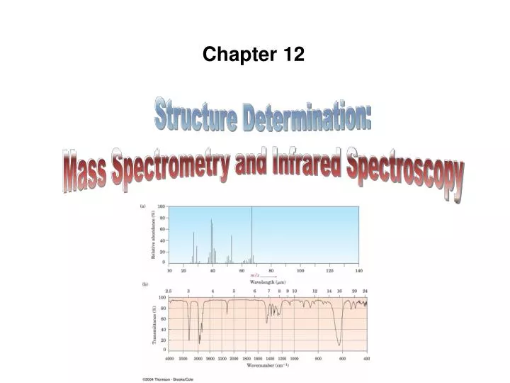
Chapter 12 Structure Determination: Mass Spectrometry and Infrared Spectroscopy
Introduction • The analysis of the outcome of a reaction requires that we know the full structure of the products as well as the reactants
Determining the Structure of an Organic Compound • In the 19th and early 20th centuries, structures were determined by synthesis and chemical degradation that related compounds to each other • Powerful techniques are now available that greatly simplify the problem of structure determination
Physical methods now permit structures to be determined directly. • We will examine: • Mass Spectrometry (MS) — this chapter • Infrared (IR) Spectroscopy — this chapter • Nuclear Magnetic Resonance Spectroscopy (NMR) —Chapter 13 • Ultraviolet Spectroscopy (UV)—Chapter 14
Mass Spectrometry (MS) – determines the size and formula • Infrared (IR) Spectroscopy– determines the kinds of functional groups present • Nuclear Magnetic Resonance Spectroscopy (NMR) –– determines the carbon- hydrogen framework • Ultraviolet Spectroscopy (UV)– determines if a conjugated p electron system is present
1.Mass Spectrometry • Mass Spectrometry (MS)– is a technique used to measure the mass, and thus the molecular weight (MW), of a molecule • It also provides structural information about a molecule from the masses of fragments produced
Mass Spectrometers have three basic parts: • an ionization source • a mass analyzer • a detector Sample Display Ionization source Mass Analyzer Detector Sample molecules are given an electrical charge Ions are separated by their mass-to- charge ratio Separated ions are observed and counted
In the ionization source, sample is vaporized and bombarded by high-energy electrons that remove an electron. • This creates a cation-radical • Cation: Molecule is positively charged (after losing an electron) • Radical: Molecule has an odd number of electrons
Bonds in cation radicals begin to break (fragment). • Some are positively charged (cations) • Some are neutral • The mass analyzer separates the ions by their mass-to-charge ratio (m/z) • Mass-to-charge ratio (m/z) is measured • The detector records the fragments as peaks at the various m/z ratios • z is usually 1. Thus, m/z is m.
Mass Spectrum • Mass spectrum – plots mass of ions (m/z) (x-axis) versus the intensity of the signal (roughly corresponding to the number of ions) (y-axis) • Tallest peak is base peak (100%) • Other peaks listed as the % of that peak • Peak that corresponds to the unfragmented radical cation is parent peak or molecular ion(M+)
MS Examples: Methane and Propane • Methane produces a parent peak (m/z = 16) and fragments of 15 and 14 (See Figure 12-2 a)
MS Examples: Methane and Propane • The MS of propane is more complex (Figure 12-2 b) since the molecule can break down in several ways
Figure 12-2 a Figure 12-2 b
2.Interpreting Mass Spectra • Mass Spectrometry (MS)– is a technique used to determine the molecular weight (MW) from the mass of the molecular ion • Double-focusingMS instruments - have such high-resolution that they provide “exact mass” • They distinguish specific atoms at an accuracy of 0.0001 atomic mass units
Double-focusing instruments provide “exact mass” • Example: MW “72” is ambiguous: • C5H12 and C4H8O have MW = 72 but: • C5H12 has an exact mass 72.0939 amu and • C4H8O has an exact mass 72.0575 amu • This is the result from fractional mass differences of atoms 16O = 15.99491, 12C = 12.0000, 1H = 1.00783 • Instruments measure the sum of the exact atomic masses of each isotope in a molecule • They include computation of formulas for each peak
Other Mass Spectral Features • Some compounds fragment so easily that no molecular ion M+ is observed on the mass spectrum • Example: 2,2-dimethylpropane (C5H12; MW = 72) No M+ is observed when electron-impact ionization is used
Other Mass Spectral Features • If molecular ion (M+) is not present due to electron bombardment causing breakdown, “softer” methods such as chemical ionization are used • Peaks above the molecular weight(M+1) appear as a result of naturally occurring heavier isotopes in the sample (i.e 13C and/or 2H) • (M+1) may be due to: • 13C that is randomly present (1.10% natural abundance) • 2H (deuterium) that is randomly present (0.15% natural abundance)
Practice Problem: Write as many molecular formulas as you can for compounds that have the following molecular ions in their mass spectra. Assume that all the compounds contain C and H and that O may or may not be present. • M+ = 86 • M+ = 128 • M+ = 156
Practice Problem: The male sex hormone testosterone contains C, H, and O and has a mass of 288.2089 amu as determined by high-resolution mass spectrometry. What is the molecular formula of testosterone?
3.Interpreting Mass SpectralFragmentation Patterns • Fragmentation pattern – is the way molecular ions break down to produce characteristic fragments that help in identification • It serves as a “fingerprint” for comparison with known materials in analysis (used in forensics) recorded in a computerized data base called the Registry of Mass Spectral data • It also provides structural clues
Fragmentation Pattern • Fragmentationoccurs when the high-energy cation radical breaks down by spontaneous cleavage of a chemical bond forming: • a carbocation (the fragment with the positive charge) • a neutral radical (the other fragment) • Positive charge goes to fragments that best can stabilize it • Stable carbocation is formed
Mass Spectral Fragmentation of Hexane • Hexane (m/z = 86 for parent) has peaks at m/z = 71, 57, 43, 29
Mass Spectral Fragmentation of Hexane • Hexane fragments as follows:
Practice Problem: methylcyclohexane or ethylcyclopentane? M+ - CH2CH3 = 69 M+ = 98 M+ - CH3 = 83 M+ = 98
Practice Problem: Two mass spectra are shown. One spectrum corresponds to 2-methyl-2-pentene; the other, to 2-hexene. Which is which? Explain. M+ - CH3 = 69 M+ = 84 M+ - CH2CH3 = 55 M+ = 84
4.Mass Spectral Behavior of Some Common Functional Groups • Mass-spectral fragmentations are usually complex and difficult to interpret. • However, there are some distinguishing features of several common functional groups • Functional groups cause common patterns of cleavage in their vicinity
Mass Spectral Cleavage Reactions of Alcohols • Alcohols undergo • alpha () cleavage (at the bond next to the C-OH) and • dehydration (loss of H-OH) to give C=C neutral radical alkene radical cation m/z = (M+ -18)
Mass Spectral Cleavage Reactions of Amines • Amines undergo • alpha () cleavage (at the bond next to the C-N), generating an alkyl radical and a N-containing cation alkyl radical
Mass Spectral Cleavage of Carbonyl compounds • Ketones and aldehydes undergo: • McLafferty rearrangement • a H on a carbon three atoms away from the carbonyl group (C=O) is transferred to the O of the C=O, • a C-C bond is broken, and • a neutral alkene fragment is produced • alpha () cleavage • at the bond between the C=O and the neighboring C
Ketones and aldehydes undergo: • McLafferty rearrangement • alpha () cleavage neutral radical
Practice Problem: Identify fragments for 2-methyl-3-pentanol MS
Practice Problem: What are the masses of the charged fragments produced in the following cleavage pathways? • Alpha cleavage of 2-pentanone (CH3COCH2CH2CH3) • Dehydration of cyclohexanol (hydroxycyclohexane) • McLafferty rearrangement of 4-methyl-2-pentanone • [CH3COCH2CH(CH3)2] • Alpha cleavage of triethylamine [(CH3CH2)3N]
Practice Problem: List the masses of the parent ion and of several fragments you might expect to find in the mass spectrum of the following molecule (red = O)
5.Spectroscopy and the Electromagnetic Spectrum • Unlike mass spectrometry, infrared (IR), ultraviolet (UV) and nuclear magnetic resonance (NMR) spectroscopies: • are nondestructive • involve interaction of molecules with electromagnetic energy rather than with high-energy electron beam
The Electromagnetic Spectrum • The electromagnetic spectrum is the range of electromagnetic energy, including IR, UV and visible radiation
The electromagnetic spectrum covers a continuous range of wavelengths and frequencies, radio waves to g rays Low n High n Low l High l
Electromagnetic radiation has dual behavior: • It behaves as a particle (called a photon) • It behaves as an energy wave • Electromagnetic energy is transmitted only in discrete amounts called quanta. • Electromagnetic waves are characterized by: • a wavelength (l) • a frequency (n) • anamplitude
Wavelength (l) –is the distance from one wave maximum to the next • Frequency (n) -is the # of waves that pass by a fixed point per unit time (s-1 or Hz) • Amplitude -is the height of a wave, measured from midpoint to peak • Wavelength x Frequency = Speed • l (m)x n (s-1)= c c c l = n= n l Speed of light: Cvacuum = 3.00 x 10⁸ m/s
hc hn • The Planck equation gives: e = = l where e = Energy of 1 photon (1 quantum) h = Planck’s constant (6.62 x10⁻³⁴J.s) n = Frequency (s-1) l = Wavelength (m) c= Speed of light (3.00 x 10⁸ m/s) • Radiant energy is proportional to its frequency and inversely proportional to its wavelength
NA hc 1.20 x 10⁻⁴ kJ/mol • The Planck equation can be rewritten: E=NAe = = l l where E = Energy of Avogadro’s number of photons NA= Avogadro’s number e = Energy of 1 photon (1 quantum) h = Planck’s constant (6.62 x10⁻³⁴J.s) c= Speed of light (3.00 x 10⁸ m/s) l = Wavelength (m)
Absorption Spectrum • Organic compounds exposed to electromagnetic radiation can absorb energy of certain wavelengths but transmit energy of other wavelengths • They can absorb photons of specific energies (wavelengths or frequencies) • Changing wavelengths to determine which are absorbed and which are transmitted produces an absorption spectrum • Energy absorbed is distributed internally in a distinct and reproducible way
An absorption spectrum shows the wavelength on the x-axis and the intensity of the various energy absorptions expressed in % transmittance on the y-axis. Ethyl alcohol CH3CH2OH
Practice Problem: Which has higher energy, infrared radiation with l = 1.0 x 10⁻⁶ m or an X ray with l = 3.0 x 10⁻⁹ m?
Practice Problem: Which has higher energy, radiation with n = 4.0 x 10⁹ Hz or radiation with l = 9.0 x 10⁻⁶ m?
Practice Problem: Calculate the energies of each of the following kinds of radiation using the relationships c . 1.20 X 10⁻⁴ kJ/mol . E = and n = l l(m) • A gamma ray with l = 5.0 x 10⁻¹¹ m • An X-ray with l = 3.0 x 10⁻⁹ m • Ultraviolet light with n =6.0 x 10¹⁵ Hz • Visible light with n =7.0 x 10¹⁴ Hz • Infrared radiation with l = 2.0 x 10⁻⁵ m • Microwave radiation with n =1.0 x 10¹¹ Hz
6.Infrared Spectroscopy of OrganicMolecules • The infrared (IR) region is lower in photon energy than visible light • Only 2.5 106 m to 2.5 105 m region is used by organic chemists for structural analysis
Absorption Spectrum • IR energy in a spectrum is usually measured as wavenumber • Wavenumber (n)is the inverse of wavelength is proportional to frequency is expressed in cm-1 ~ 1 Wavenumber(cm-1)= l (cm) • Specific IR absorbed by organic molecule is related to its structure