Download
1 / 84
840 likes | 987 Views
Respiratory System. Respiration. Ventilation : Movement of air into and out of lungs External respiration : Gas exchange between air in lungs and blood Transport of oxygen and carbon dioxide in the blood Internal respiration : Gas exchange between the blood and tissues.

E N D
Respiration • Ventilation: Movement of air into and out of lungs • External respiration: Gas exchange between air in lungs and blood • Transport of oxygen and carbon dioxide in the blood • Internal respiration: Gas exchange between the blood and tissues
Respiratory System Functions • Gas exchange: Oxygen enters blood and carbon dioxide leaves • Regulation of blood pH: Altered by changing blood carbon dioxide levels • Voice production: Movement of air past vocal folds makes sound and speech • Olfaction: Smell occurs when airborne molecules drawn into nasal cavity • Protection: Against microorganisms by preventing entry and removing them
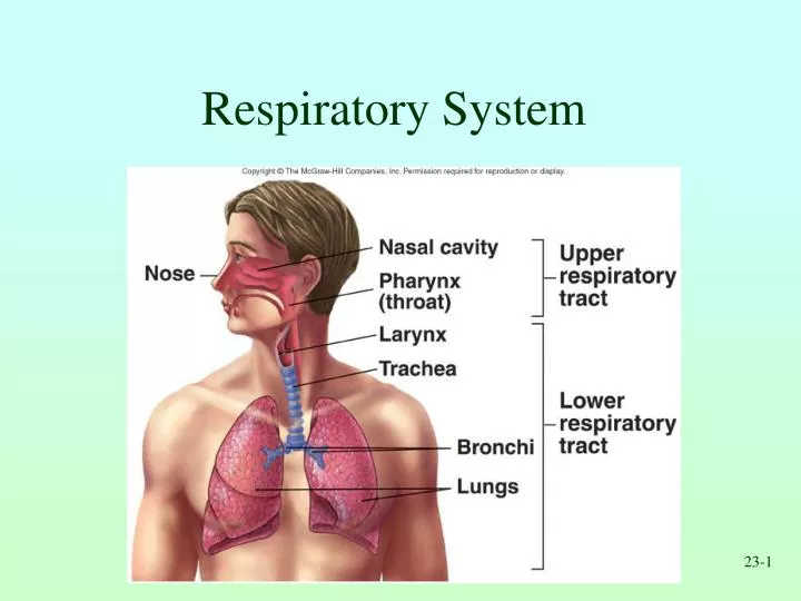
Respiratory System Divisions • Upper tract • Nose, pharynx and associated structures • Lower tract • Larynx, trachea, bronchi, lungs
Nose External nose Nasal cavity Functions Passageway for air Cleans the air Humidifies, warms air Smell Along with paranasal sinuses are resonating chambers for speech Pharynx Common opening for digestive and respiratory systems Three regions Nasopharynx Oropharynx Laryngopharynx Nose and Pharynx
Larynx • Functions • Maintain an open passageway for air movement • Epiglottis and vestibular folds prevent swallowed material from moving into larynx • Vocal folds are primary source of sound production
Trachea • Windpipe • Divides to form • Primary bronchi Insert Fig 23.5 all but b
Tracheobronchial Tree • Conducting zone • Trachea to terminal bronchioles which is ciliated for removal of debris • Passageway for air movement • Cartilage holds tube system open and smooth muscle controls tube diameter • Respiratory zone • Respiratory bronchioles to alveoli • Site for gas exchange
Lungs • Two lungs: Principal organs of respiration • Right lung: Three lobes • Left lung: Two lobes • Divisions • Lobes, bronchopulmonary segments, lobules
Ventilation • Movement of air into and out of lungs • Air moves from area of higher pressure to area of lower pressure • Pressure is inversely related to volume
Basic Chest X-Ray Interpretation Deb Updegraff, C.N.S., PICU
X-rays- describe radiation which is part of the spectrum which includes visible light, gamma rays and cosmic radiation. Unlike visible light, radiation passes through stuff. When you shine a beam of X-Ray at a person and put a film on the other side of them a shadow is produced of the inside of their body.
Different tissues in our body absorb X-rays at different extents: • Bone- high absorption (white) • Tissue- somewhere in the middle absorption (grey) • Air- low absorption (black)
Film Quality • First determine is the film a PA or AP view. PA- the x-rays penetrate through the back of the patient on to the film AP-the x-rays penetrate through the front of the patient on to the film. All x-rays in the PICU are portable and are AP view
Quality (cont.) • Is the film over or under penetrated if under penetrated you will not be able to see the thoracic vertebrae.
Quality (cont) • Check for rotation • Does the thoracic spine align in the center of the sternum and between the clavicles? • Are the clavicles level?
LUNG VOLUMES • The total volume contained in the lung at the end of a maximal inspiration is subdivided into volumes and subdivided into capacities. • There are four volume subdivisions which: • do not overlap. • can not be further divided. • when added together equal total lung capacity.
Capacities • Lung capacities are subdivisions of total volume that include two or more of the 4 basic lung volumes.
Basic lung volumes (memorize) • Tidal Volume (TV). The amount of gas inspired or expired with each breath. • Inspiratory Reserve Volume (IRV). Maximum amount of additional air that can be inspired from the end of a normal inspiration.
Basic lung volumes (memorize) • Expiratory Reserve Volume (ERV). The maximum volume of additional air that can be expired from the end of a normal expiration. • Residual Volume (RV). The volume of air remaining in the lung after a maximal expiration. This is the only lung volume which cannot be measured with a spirometer.
Basic lung capacities (memorize) • Total Lung Capacity (TLC). The volume of air contained in the lungs at the end of a maximal inspiration. Called a capacity because it is the sum of the 4 basic lung volumes. TLC=RV+IRV+TV+ERV
Basic lung capacities (memorize) • Vital Capacity (VC). The maximum volume of air that can be forcefully expelled from the lungs following a maximal inspiration. Called a capacity because it is the sum of inspiratory reserve volume, tidal volume, and expiratory reserve volume. VC=IRV+TV+ERV=TLC-RV
Basic lung capacities (memorize) • Functional Residual Capacity (FRC). The volume of air remaining in the lung at the end of a normal expiration. Called a capacity because it equals residual volume plus expiratory reserve volume. FRC=RV+ERV
Basic lung capacities (memorize) • Inspiratory Capacity (IC). Maximum volume of air that can be inspired from end expiratory position. Called a capacity because it is the sum of tidal volume and inspiratory reserve volume. This capacity is of less clinical significance than the other three. IC=TV+IRV
Now you are ready • Look at the diaphram: for tenting free air abnormal elevation • Margins should be sharp (the right hemidiaphram is usually slightly higher than the left)
Check the Heart • Size • Shape • Silhouette-margins should be sharp • Diameter (>1/2 thoracic diameter is enlarged heart) Remember: AP views make heart appear larger than it actually is.
Cardiac Silhouette • R Atrium • R Ventricle • 3. Apex of L Ventricle • Superior Vena Cava • Inferior Vena Cava • 6. Tricuspid Valve • Pulmonary Valve • Pulmonary Trunk • 9. R PA 10. L PA
Check the costophrenic angles Margins should be sharp
Check the hilar region • The hilar – the large blood vessels going to and from the lung at the root of each lung where it meets the heart. • Check for size and shape of aorta, nodes,enlarged vessels
Finally, Check the Lung Fields • Infiltrates • Increased interstitial markings • Masses • Absence of normal margins • Air bronchograms • Increased vascularity