Download
1 / 43
500 likes | 1.1k Views
Pathological Diagnosis of Mycoplasmosis in Swine. Swine Mycoplasma Pneumonia Workshop FDA / CVM March 6,7 of 2002 Kansas City, MO Kent Schwartz. Swine Pneumonia: Early Years. “Normal” for pigs to cough / scratch Enzootic (Mycoplasma + Pasteurella) Ascarid Migration

E N D
Pathological Diagnosis of Mycoplasmosis in Swine Swine Mycoplasma Pneumonia Workshop FDA / CVM March 6,7 of 2002 Kansas City, MO Kent Schwartz
Swine Pneumonia: Early Years • “Normal” for pigs to cough / scratch • Enzootic (Mycoplasma + Pasteurella) • Ascarid Migration • 1918: H1N1 Swine Influenza • Atrophic rhinitis (Bordetella and Pasteurella) • Small farms / “home remedies”
Mechanization: Trend to Confinement and Larger Herds • M hyo, SIV, ascarids, AR • More science, agents, diagnostics • PRV, Actinobacillus pleuropneumonia • Pasteurella multocida with M hyo • Age of therapeutics (antimicrobials) • “Vaccination” products
Era of Altered Ecology/New Agents • Segregated rearing / larger populations / Altered herd immunity / altered ecology • M hyo, SIV, App remain • Bacterial “Opportunists” Emerge • Hemophilus parasuis, Streptococcus suis • Actinobacillus suis, Salmonella sp. others • “New” agents; respiratory and systemic • PRRSV, PCV, SIV H3N2, PRCV • Porcine Respiratory Disease Complex (PRDC) is a multifactorial culmination
“Immune Confusion” • Respiratory Tract: mixing vessel for: • Systemic diseases (PRRSV, PCV, bacteria) • Respiratory agents (SIV, bacteria) • M hyo • “Herd immunity” is variable for agents • Sow herd “stability” influences maternal immunity • Piglet infection status variable and sequential • Immune status is variable • Many permutations of agents involved • Many permutations in sequence of infections and sequence may matter
M hyo remains Central and Primary • Infection is common; not easily eliminated • Infection persists for months • Alters mechanical clearance of debris • Inflammation / immune-mediated damage • Altered, nonproductive immune response • “Immunologically privileged” site of infection so clearance is compromised • Provides sites for opportunists • Synergy with other lung pathogens
Clinical Disease: M hyo alone • Mild malaise • No fever • Cough: nonproductive / chronic • Moderate morbidity / no mortality • Altered growth performance • ADG • Feed efficiency
Enzootic pneumonia=M hyo + bacteria • M hyo facilitates bacterial infections • Cough, malaise and anorexia • Moderate fevers 1030-1050 • Expiratory dyspnea / “thump” • Variable morbidity and mortality • Stunting, chronic pneumonia, death • Strategic interventions (medication or vaccination) can influence outcome and/or subsequent disease severity
PRDC = Enzootic pneumonia + virus(M hyo + bacteria + virus) • Severe depression, high fever, anorexia • Expiratory dyspnea (thump) • Rapid loss of condition • Medication less efficacious • High morbidity and frequently high mortality (5-20%)
Gross Pathology: • Bronchopneumonia • Cranioventral, firm, exudate in airways • Interstitial pneumonia • Diffuse, mottled, lobular distribution • Edema fluid in airways • Both can occur simultaneously: common in field cases of PRDC
M hyo: Gross Diagnosis • Early: 10 days-4 weeks • Cranioventral, lobular, red, firmness • Active: 2-6 weeks • Clearly demarcated, grayish, atelectasis • Airways prominent with mucopus • Resolution: 5-20 weeks • Gray fissures of atelectasis • Distorted lobe structure of normal lung tissue • A population will have animals at all stages of disease with variable severity
Histopathology: Basics • Bronchopneumonia • Extends from small airways • Bronchiolitis with exudate and debris • Adjacent alveoli, interstitium • Interstitial pneumonia • extends from alveolar septae • can involve small airways • Both often present in chronic disease or in mixed infections.
Histopathology is Fallible • Agents and agent combinations outnumber possible responses • Chronic lesions are less specific • “Classic” lesions only with single agents at some stages of disease • Few lesions are pathognomonic or “etiologically specific” • “Lesions are compatible with….”
M hyo: Histopathological Diagnosis • Early: 10 days-4 weeks • peribronchiolar lymphohistiocytic inflammation; scattered neutrophils • Active: 2-12 weeks • mucopus, atelectasis • Resolution: 5-20 weeks • BALT hyperplasia • A population will have animals at all stages of disease with variation in lesion severity
What else can “look” like M hyo ? • Chronic persistence of antigen/agent • Lymphoid hyperplasia / airway cuffs • Subacute SIV = Early M hyo • Ascarid migration; resolving • Chronic bacterial pneumonia • Chronic viral pneumonia • It is often difficult to demonstrate organisms in chronic lesions
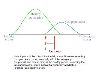
Key Points: Pathological Diagnosis • Gross lesions are “compatible with but not specific for” M hyo • Microscopic lesions are “compatible with but are not specific for” M hyo • Sensitivity and Specificity of pathology?? • Most field cases are mixed infections • M hyo, bacteria, viruses • Time for resolution varies with: • Severity and extent of initial lesion • Presence of concurrent pathogens
Etiologic diagnosis requires: • Demonstration of specific agent • M hyo isolation is not routine • Demonstration of specific antigen • IHC, FAT, ELISA • Demonstration of specific agent nucleic acid • PCR and PCR based assays • Serology confirms antibody but is NOT a definitive etiologic diagnosis
Diagnosis of Swine Pneumonia • Research / Infection models • controlled infection, controlled specimen collection, and standardized evaluations • Predictable outcomes / valid measures • Field Cases: variability is uncontrolled • Descriptive pathology has limitations • Demonstrate agents: specimen dependant • age, stage, specimens, interventions • Interpret results in the context of clinical signs, history and population dynamics
An accurate and useful field diagnosis uses all available information • Clinical signs, history, diagnostic records • Production records • Compatible gross lesions • Compatible microscopic lesions • Identify agents with appropriate tests • Primary agents, secondary agents • Define epidemiology in the population • Serologic: cross-sectional or longitudinal • In population, infection and immunity status varies so need statistical sampling techniques
Mixed agents, duration, population variability makes definitive diagnosis a challenge
Summary: M hyo in swine • M hyo is common in swine populations • M hyo alone is mild disease with cough, suppression of growth and feed efficiency • M hyo duration of effect (3-20 weeks) creates opportunities for co-infections • Not all are affected equally/simultaneously • Enzootic pneumonia is M hyo + bacteria • PRDC is M hyo + bacteria + viruses
Summary: M hyo in swine • Takes time for lesions to develop / resolve • Pathology is useful to describe severity • Variability of lesion severity in populations • Most field cases are mixed infections • Field measures of current interventions • Reduced prevalence of clinical pneumonia • Reduced lesion prevalence and severity • Reduced medication cost, treatments • Less variation in growth rate • Control of M hyo disease severity often does mitigate severity of other endemic diseases • Infection models are useful to evaluate M hyo intervention strategies and disease interactions