Download
1 / 15
160 likes | 217 Views
Explore the intricate details of the knee joint complex, including bone structure, ligaments, musculature, and common injuries. Understand the dynamic stability system that supports this crucial joint in the body.

E N D
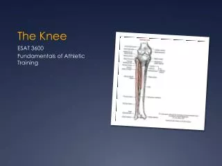
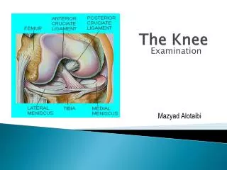
The Knee Anatomy Mazyad Alotaibi
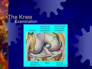
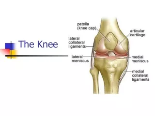
The Knee Joint Complex • Tibiofemoral Joint* • Patellofemoral Joint* • Tibiofibular Joint • Capsular Pattern – Greater loss of flexion than extension
Bones • Femur – condyles covered with articular cartilage, intercondylar groove, patella groove • Tibia – bifid plateau • Patella – medial and lateral articulating facets -increases the lever arm of the quadriceps -increases the distribution of compressive force on the femur in full flexion
Ligaments Strong ligaments provide the static stability system for the knee
MCL & LCL • Provide medial/ lateral stability and prevent excessive external rotation of the tibia • Frequently injured on the joint line • MCL – Frequently injured by an external rotation strain • Has deep and superficial fibres • LCL – Frequently injured by adduction blow to the knee
ACL • Ant part of intercondylar area • Up, back, lateral • Med asp of lat fem condyle
ACL & PCL • Provide ant / post stability of the knee • ACL – also controls rotation • Commonly injured by forced internal rotation of the femur on a fixed tibia and flexed knee • PCL – strongest -Commonly injured in flexion with an anterior force
Joint capsule – attached to the medial meniscus and MCL • Coronary Ligaments – bind the menisci to the tibia
Menisci • Lateral and medial • Peripherally thicker than central • Transverse lig attaches • Increase stability • Shock absorbing • Lubrication and nutrition • Injured during twisting activities
ITB – anterior and lateral stability and prevent excessive internal rotation of the tibia • Quadriceps – Lat vs med to maintain patella in groove • Sartorius and Gracilis – medial stability, knee and hip flex
Hamstrings • Prevent anterior displacement of the tibia • Pes anserine – sartorius, gracilis and semitendinosis. • Biceps femoris • Primarily produce knee flex and also external rotation of the tibia
Gastrocnemius – primarily ankle flexor also assists with knee flexion • Popliteus – WB external rotation and extension of the tibia • Attaches to the posterior horn of the lateral meniscus and pulls back the posterior horn to unlock the knee
Bursa • Suprapatellar – continuation of the synovial sac • Prepatellar • Deep and superficial infrapatellar • Also semimembranosus and med head of gastroc, • Gastroc heads and capsule • Pes anserine tendon
Surface markings • Joint line • Medial collateral ligament • Lateral collateral ligament • Medial coronarys • Gerdy’s Tubercle • Pes Anseurine Bursa