Download
1 / 37
370 likes | 385 Views
Learn about the development of the immune system, gestational tolerance, preventing rejection, and fetal/neonatal protection. Discover the importance of vaccinations and immunization from birth to adolescence.

E N D
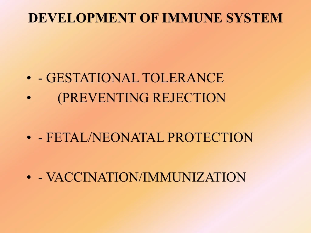
DEVELOPMENT OF IMMUNE SYSTEM • - GESTATIONAL TOLERANCE • (PREVENTING REJECTION • - FETAL/NEONATAL PROTECTION • - VACCINATION/IMMUNIZATION
BIRTH BCG (BACILLUS CALMETTE-GUERIN) ORAL POLIO HEPATITIS 6 WEEKS DPT (DIPHTHERIA, TETANUS, PERTUSSIS ORAL POLIO 2ND DOSE HEPATITIS 2ND 10 WEEKS DPT (DIPHTHERIA, TETANUS, PERTUSSIS) ORAL POLIO 3RD 14 WEEKS DPT 3RD ORAL POLIO 4TH 6-9 MONTHS ORAL POLIO 5TH HEPATITIS B 9 MONTHS MEASLES 15-18 MONTHS MMR (MEASLES, MUMPS, RUBELLA) DPT booster dose ORAL POLIO 6TH 5 YEARS DPT 2ND booster ORAL POLIO 7TH 10 YEARS TT (TETANUS) 3RD booster HEPATITIS B booster 15-16 YEARS TETANUS booster VACCINATIONS
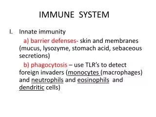
Function of Immune System is PROTECTION against: • Bacteria • Virus • Fungus/ multicellular parasites • Cancer • Toxins • ( 5,000 daltons--protein/lipid/CHO/nucleic acids)
Tissues and Organs Important for Immune Function • Cells derived from stem cells: liver, bone marrow • Cells are stored, multiply, interact, and mature in: thymus, spleen, lymph nodes, blood • Transport: lymphatic vessels • Accessory Organs • Appendix, tonsils, intestines
Cell Types • Lymphocytes: derived in bone marrow from stem cells 10^12 • A) T cells: stored & mature in thymus-migrate throughout the body • -Killer Cells • Perform lysis (infected cells) • Cell mediated immune response -Helper Cells Enhance T killer or B cell activity -Supressor Cells Reduce/suppress immune activity May help prevent auto immune disease
Lymphocytes (cont.) • B-Cells: stored and mature in spleen • secrete highly specific Ab to bind foreign substance (antigen: Ag), form Ab-Ag complex • responsible for humoral response • perform antigen processing and presentation • differentiate into plasma cells (large Ab secretion)
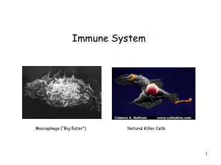
Neutrophils- found throughout body, in blood -phagocytosis of Ab-Ag CX Macrophages- throughout body, blood, lymphatics -phagocytose non-specifically (non Ab coated Ag) -phagocytose specifically Ab-Ag CX -have large number of lysosomes (degradative enzyme) -perform Ag processing and presentation -present Ag to T helper cell -secrete lymphokines/ cytokines to stimulate T helper cells and immune activity 4. Natural Killer Cells-in blood throughout body -destroy cancer cells -stimulated by interferons
Macrophage Bacteria Bacterial Infection
Complement Series of enzymes which are sequentially activated and result in lysis of cell membrane of infected cell at bacterium Permeablizes membrane leaky Complement binding and activation ~35 enzymes and factors involved in cascade
5 classes of Ig IgG: 150,000 m.w. most abundant in blood, cross placental barrier, fix complement, induce macrophage engulfment IgA: associated with mucus and secretory glands, respiratory tract, intestines, saliva, tears, milk variable size IgM: 900,000 m.w. 2nd most abundant , fix complement, induce macrophage engulfment, primary immune response
5 Classes of Ig IgD: Low level in blood, surface receptor on B- cell IgE: Binds receptor on mast cells (basophils) secretes histamine, role in allergic reactions Increased histamine leads to vasodilation, which leads to increase blood vessel permeability. This induces lymphocyte immigration swelling and redness.
Thymus Involution Repertoire of lymphocytes shift with aging (membrane components shift)
ORGAN AND T-CELL DEVELOPMENT • YOLK SAC • LIVER • (4 Weeks) • BONE MARROW • (4-5 Weeks ) • THYMUS • (7-10 Weeks) • BLOOD LYMPH • (14 Weeks) • SPLEEN • (16 Weeks) • T-cells migrate and appear in tissues with development and increase in number throughout Gestation
B-CELLS • FIRST appear in immature state - Liver at 7 weeks • LATER –appear mature by 14-20 weeks • CAN DIFFERENTIATE INTO IMMUNOLOGICALLY COMPETENT ANTIBODY-PRODUCING PLASMA CELLS
NATURAL KILLER CELLS • FIRST APPEAR IN FETAL BONE MARROW AROUND 13 WEEKS GESTATION • FIRST APPEAR IN FETAL BONE MARROW AROUND 13 WEEKS GESTATION • FOUND THROUGHOUT BODY • NK CELLS HAVE DIMINISHED ACTIVITY BEFORE BIRTH COMPARED TO ADULT • STIMULATED BY INTERFERON AFTER 27 WEEKS
COMPLEMENT PROTEINS • ARISE FROM LIVER • FIRST DETECTED 5-6 WEEKS GESTATION • INCREASE GRADUALLY IN CONCENTRATION • AT ABOUT 28 WEEKS COMPLEMENT PROTEINS ARE AROUND 2/3 THAT OF ADULT CONCENTRATIONS • INDIVIDUAL VARIATION
SEVERE COMBINED IMMUNODEFICIENCY DISEASE (SCID) CHARACTERISTICS: GENERALLY CAUSED BY DEFECT OF SINGLE GENE NEEDED FOR T-CELL AND B-CELL FUNCTION —SUBJECT EXHIBITS NO CELL MEDIATED RESPONSE ––SUBJECT CANNOT MAKE ANTIBODIES ABOUT 25% OF CASES INVOLVES DEFECTIVE GENE FOR THE ENZYME ADENOSINE DEAMINASE (REQUIRED FOR PURINE BREAKDOWN)
SEVERE COMBINED IMMUNODEFICIENCY DISEASE (SCID) • TREATMENT OPTIONS: • GERM FREE ENVIRONMENT • BONE MARROW TRANSPLANT • ROUTINE INJECTIONS OF ADENOSINE DEAMINASE ENZYME (ADA) • GENE THERAPY USING SUBJECTS OWN CELLS • (RETROVIRUS CONTAINING ADA TO “INFECT” • SUBJECTS BONE MARROW STEM CELLS)
TABLE 15.6 - CHEMICAL MEDIATORS OR MODULATORS CYTOKINES - influence proliferation, differentiation, and survival of lymphoid cells; has numerous actions on other body cells, compromises the following: Granulocyte-Macrophage Colony Stimulating Factor (GM-CSF):regulates hematopoiesis, affects phagocyte function and angiogenesis Interleukin (IL):family, 16 different proteins from IL1 and up; numerous effects on lymphocytes and other cells with IL receptors Colony Stimulating Factor (CSF):glycoprotein regulating white blood cell production, activity, and survival Tumor Necrosis Factor (TNF):1) TNF- cytotoxic against malignant and inflammatory cells; produced primarily by macrophages, 2) TNF- cytotoxic against malignant cells; enhances phagocytosis; produced primarily by T cells Interferon (IFN): 1) IFN-,: produced by many cells; antiviral actions, 2) IFN-:synthesized by activated NK and T cells; involved in activation of macrophages and inflammation
Experimental Evidence for Age Related Decrease in Immune Function Sheep RBC (Antigen) 1st into human Dependent on T & B cell function
Table 15-2: Some Aging Related Effects on B-Cells • Decreased number of circulating and peripheral blood B cells • Alteration in B-cell repertoire (diversity) • Decreased generation of primary and secondary memory B cells • General decline in lymphoproliferative capacity
Table 15-14: Some Aging-Related Effects on T-cells • General decline in cell mediated immunological function • T-cell population is hyporesponsive • Decrease responsiveness in T-cell repertoire (i.e. diversity of CD8+ T-cells) • Decline in new T-cell production • Increase in proportion of memory and activated T-cells while naïve T-cells decrease • Diminished functional capacity of naïve T-cells (decreased proliferation, survival, and IL-2 production) • Senescent T-cells accumulate due to defects in apoptosis • Increased proportion of thymocytes with immature phenotype • Shift in lymphocyte population from T-cells to NK/T cells (cell expressing both T-cell receptor and NK cell receptors)
Table 15-13 Aging-Related Shifts in Antibodies • General decrease in humoral responsiveness: • Decline in high affinity protective antibody production • Increased auto-antibodies: • Organ specific and non-organ specific • antibodies directed to self • Increased serum levels of IgG (i.e. IgG1 and IgG3) and IgA; IgM levels remain unchanged
Table 15-16 Influence of Aging on Macrophages • and Granulocytes • General functional impairment of macrophages and granulocytes • GM-CSF is unable to activate granulocytes from elderly subjects (e.g.: superoxide production and cytotoxic abilities) • Polymorphonuclear neutrophils appear to possess higher levels of surface markers CD15 and CD11b and lesser vesicles containing CD69 which lead to the impairment observed to destroy a bacteria • In elderly subjects the monocyte phenotype shifts (i.e. expansion of CD14dim and CD16 bright subpopulations which have features in common with mature tissue macrophages) • Macrophages of aged mice may produce less IFN-, less nitric oxide synthetase, and hydrogen peroxide.
Table 15-15 Aging-Related Changes in Natural Killer (NK) Cells • General decline in cell function • Good correlation between mortality risk and NK cell number • Increased in proportion of cells with high NK activity (i.e. CD16+, CD57-) • Progressive increase in percentage of NK cells • Impairment of cytotoxic capacity per NK cell • Increase in NK cells having surface molecule CD56 • dim subset
Table 15-10 Some Aging-Related Shifts in Cytokines • Increased proinflammatory cytokines IL-1, IL-6, TNF- • Increased cytokine production imbalance • Decreased IL-2 production • Increased production of IL-8, which can recruit macrophages and may lead to pulmonary inflammation • Increase in dysfunctional IL-8 • Decreased secretion of IFN- (interferon) • Altered cytokine responsiveness of NK cells, which have decreased functional abilities • Increased levels of IL-10 and IL-12 upregulated by Antigen Processing Cells
Table 15-17 Major Diseases Associated with Aging • in Immune Function • Increased tumor incidence and cancer • Increased incidence of infectious diseases caused by: • E. Coli • Streptococcus pneumonia • Mycobacterium tuberculosis • Pseudomonas aeruginosa • Herpes virus • Gastroenteritis, bronchitis, and influenza • Reappearance of latent viral infection • Autoimmune diseases and inflammatory reactions: • Arthritis • Diabetes • Osteoporosis • Dementia
Table 15-9 Hallmarks of Immunosenescence • Atrophy of the thymus: • decreased size • decreased cellularity (fewer thymocytes and epithelial cells) • morphologic disorganization • Decline in the production of new cells from the bone marrow • Decline in the number of cells exported by the thymus gland • Decline in responsiveness to vaccines • Reduction in formation and reactivity of germinal center nodules in lymph nodes where B-cells proliferate • Decreased immune surveillance by T lymphocytes and NK cells
Aging of the Immune System Dr. Hal Sternberg BioTime, Inc. Berkeley, CA
Evidence for Decline in Immune Function with Aging Aged Individuals have: 1) Increased incidence of INFECTIONS: For example: pneumonia, influenza, tuberculosis, meningitis, urinary tract infections 2) Increased incidence of AUTOIMMUNE DISEASE: For example: rheumatoid arthritis, lupus, hepatitis, thyroiditis (graves-hyper/hashimotos-hypo), multiple sclerosis (Predisposition toward these diseases is related to Human Leukocyte Antigens HLA genes)
Evidence for Decline in Immune Function with Aging Aged Individuals have: 3) Increased CANCER INCIDENCE: For Example: prostate, breast, lung, throat/neck/head, stomach/colon/bladder, skin, leukemia, pancreatic 4) TOLERANCE to organ transplants: Kidneys, skin, bone marrow, heart (valves), liver, pancreas, lungs
B-Cell Mitogenesis Strain dependent, mitogen dependent, etc. • Mitogen • Lipopolysac. • PHA In vitro
15-11 Additional Aging-Related Shifts in Immune Functions • Altered membrane fluidity • Increased apoptosis perhaps due to decline • in CD28 expression and IL-2 production • CD20 overexpression on lymphocytes • Increased CAMs expression on lymphocytes • Old cells may have greater levels of • messenger RNA for 3 mitotic inhibitors • Decrease number of HLA class I and II • antigenic sites on lymphocytes • Increase in activated T-cell expressing DR molecules • Decreased proportion of T, B, and NK cells • expressing CD62L and increased density per cell • of this adhesion receptor expression • Upregulation of L-selectin per T-cell • Shift in lymphocyte population to contain • more CD3-NK cells and CD3+CD56+ T-cells • CD3 downregulation and CD50 upregulation • on T-cells affecting activation and proliferation • Increased T-cell death by fas/fas-ligand mediated • response in presence of IL-2 • Heightened density of CD5 on B-cells • Decreased number of monocytes with LFA-1 • Decreased ability of dendritic cells to stimulate • T-cell secretion of IFN- and IL2