Download
1 / 32
370 likes | 803 Views
Immune System . Chapter 21. Immune System. Functional body system Structures are cells not organs Provides immunity Recognizes ‘self’ from ‘non-self’ Fights pathogens and infections Destroy cancer cells Isolate and remove foreign substances Divisions Innate immunity (non-specific)

E N D
Immune System Chapter 21
Immune System • Functional body system • Structures are cells not organs • Provides immunity • Recognizes ‘self’ from ‘non-self’ • Fights pathogens and infections • Destroy cancer cells • Isolate and remove foreign substances • Divisions • Innate immunity (non-specific) • Adaptive immunity (specific)
Innate Immunity • External defenses prevent entry • Skin • Mucous membranes • Internal defenses prevent/inhibit spread • Identified by surface carbs or proteins • Types • Phagocytes and natural killer (NK) cells • Inflammation (chemicals) and fever • Antimicrobial proteins
External Defenses • Most efficient when unbroken/uninjured • Skin • Keratinized stratified squamous • Mucous membranes (chemical barriers) • Lines body cavities w/external openings • Produce wide range of chemicals • Sebaceous and sweat glands • Gastric juices, urine, and vaginal secretions • Salivary and lacrimal lysozyme secretions • Nasal cilia • Table 21.2
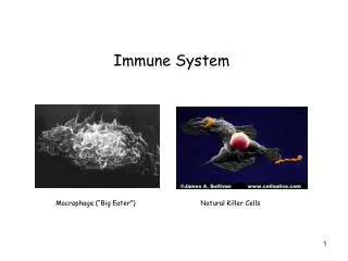
Phagocytes • Macrophages (monocytes) • Wander through tissues (free) • Kupffer cells in liver and microglia in brain (fixed) • Resilient fighters • Neutrophils • Need exposure to infectious substances • Self sacrifice fighters • Eosinophils • Against parasitic worms • Mast cells • Local inflammatory response w/ pathogen exposure (allergies) • Release histamine, heparin, and proteases
Phagocytosis • Microbe is engulfed phagosome • Phagocyte adheres to PM identifiers • Complement proteins and antibodies assist opsonization • Phagosome fuses to lysosome phagolysosome • Digestion • Respiratory burst needed for complex microbes • Helper T cells stimulate macrophages to produce • Increase pH to activate additional enzymes • Neutrophils produce defensinsto pierce PM • Exocytosis • Fig 21.2
Natural Killer (NK) Cells • Large, granular lymphocytes • Recognize and attack any ‘non-self’ cells (non-specific) • Cancer and viral cells • Perforins secreted to puncture PM • Induce apoptosis, programmed cell death • Enhance inflammatory response
Inflammatory Response • Clean up area and isolate/stop spread • Process • Histamine released by damaged tissues • Local vessel(s) vasodilation and permeability increase • Hyperema from increased blood flow (Redness and heat) • Clotting factors & antibodies (exudate) into tissues (edema) • Swelling presses on nerves releasing prostoglandins (pain) • Antihistamines and aspirin/acetaminophen reduces • Phagocytes attracted • Fig 21.3
Phagocyte Attraction • Leukocytosis • Chemical signals increase neutrophil number • Margination • Neutrophils cling to capillary walls in injured area • Cell adhesion molecules (CAM’s) signal location and facilitate attachment • Diapedesis • Neutrophils move from blood to tissue • Chemotaxis • Attracts WBC’s to area phagocytosis • Monocytes follow neutrophils macrophages • Pus with bad infections
Fever • Systemic response to pathogen invasion • Leukocytes & macrophages release pyrogens to signal hypothalamus • High is dangerous • Denaturation of proteins (enzymes) • Moderate can be beneficial • Metabolic rate up = tissue repair rate up • Liver & spleen withhold iron & zinc = starves microbe
Antimicrobial Proteins: Interferons • Synthesized by infected cells • Enter neighboring cells to ‘interfere’ w/ viral reproduction • Not virus specific • Activate macrophages and NK cells too
Antimicrobial Proteins: Complement • ‘Complements’ innate and adaptive defenses • 20+ inactive blood proteins • Activation • Classical pathway: activates through antigen/antibody binding • Alternate pathway: proteins directly attach to antigen • Result • Lyse many cell types (‘self’ are protected) • Protein complex produces a pore in PM • H2O floods in • Amplifies inflammatory response • Opsonization
Adaptive Defenses • Attacks specific foreign substances • Longer reaction (antigen exposure) • Systemic protection (blood stream residence) • Permanent protection (‘memory’) • Mechanisms • Humoral immunity utilizes antibodies • Cellular immunity utilizes lymphocytes and other phagocytes
Antigens • Molecules/cells eliciting adaptive immune response • Antigenic determinants provide signal • Immunogenicity: stimulates lymphocyte and antibody production • Reactivity: react with lymphocytes and antibodies • Types • Complete • Biological macromolecules, pollen, and microorganisms • Incomplete (haptens) • Smaller molecules that bind to ‘self’ selves • Cause hypersensitivity or allergies
MHC Proteins • Basis for ‘self’ and ‘non-self’ identification • Major histocompatibility complex genes encode • Unique to all individuals • Synthesized in the ER and transported to the PM for display • Class I on all body cells • Class II on dendritic cells, macrophages, and B-cells
Lymphocytes (revisited) • Produced in red bone marrow from hemocytoblasts • Develop immunocompetence in primary lymphoid organs • B-cells in bone marrow and T-cells in thymus • Multiple antigen receptors • Migrate to secondary lymphatic organs • Antigen binding completes differentiation • Learn self-tolerance through positive & negative selection • Effector or memory cells free to wander
Antigen-Presenting Cells (APCs) • Dendritic cells, macrophages, and B-cells • Functions • Engulf antigens for T-cell presentation • Fragments join MHC proteins on PM surface • Enables visualization of antigen by T-cell • Self/non-self complex recognized by T-cell • Binding signals differentiation • Key to initiating adaptive immunity
Humoral Immune Response • Antigen challenge when B-cell first meets antigen • Clonal selection forms … • Plasma cells antibodies circulate to ‘flag’ antigens • Memory cells quicker repeat response • Occurs in spleen or lymph nodes
Immunological Memory • Primary immune response • Antigen challenge • Secondary immune response • Stronger, faster, & longer
Humoral Immunity 2.0 • Active immunity • B-cells encounter antigens and MAKES antibodies • Natural exposure causes symptoms & suffering • Artificial exposure from dead or attenuated (vaccines) • Examples of each? • Passive immunity • Antibodies WITHOUT antigen exposure • Naturally b/w mother and fetus/infant • Artificially through pre- or post-injection • Examples of each?
Antibodies • Produce by B-cells AFTER antigen challenge • Also known as immunoglobins (Igs) • 4 looping polypeptides • Identical light (2) and heavy (2) chains • Variable regions (2 per) • Bind antigen • Constant regions • Basis for class distinction (5) • Determine functioning • Functions • Recognize and bind antigens • Inactivation of antigens Image from Purves et al., Life: The Science of Biology, 4th Edition, by Sinauer Associates (www.sinauer.com) and WH Freeman (www.whfreeman.com)
Antibody Classes • IgM • Produced 1st by plasma cells; activates complement • IgA • Prevents pathogen attachment in mucus membranes • IgD • B-cell surface receptor to activate other B-cells • IgG • Most abundant for 1° and 2° responses; fetal immunity, activates complement • IgE • Responsible for allergies due to histamine release
Targeting Antigens • Precipitation • Phagocyte accessibility increase due to weight increase • Lysis (by complement) • Antibodies (which?) insert MAC into PM (earlier) • Enhance inflammatory response and opsonization • Agglutination • Clumps antigens for phagocytes • Neutralization • Bind/block attachment and exotoxin sites on antigen • Slows until phagocytosis
Cell-Mediated Immune Response • Works when cells are infected w/antigen • Requires ‘visualization’ of antigen for T-cell • T-cells distinguished by glycoprotein receptors • CD8 cells • Recognize class I MHC proteins (all body cells) • Become cytotoxic/killer T-cells (TC) • CD4 cells • Recognize class II MHC proteins (APCs) • Become helper T-cells (TH) or memory T-cells
T-Cell Types • Cytotoxic T-cells directly attacks and kills • Perforins or apoptosis signal to ‘non-self’ cells • Helper T-cells elicit immune responses • Stimulate B-cells to proliferate • Activate CD8 cells w/ co-stimulatory chemicals • Increase macrophage strength, cytokine release, & amplify innate defenses • Memory T-cells remain to prepare for future exposure • Regulatory T-cells (TReg) inhibit immune response • Stops once unnecessary • Autoimmune disease protection
T-Cell Differentiation and Cloning • Differentiation requires double recognition • Self and non-self • Class I MHC proteins with endogenous antigens • Class II MHC proteins with exogenous antigens • CD8 and CD4 recognize and bind accordingly • Co-stimulation signals clones to be produced • Additional T-cell binding or cytokine/interleuken release • Lack of causes T-cell tolerance and prevents mitosis
Organ Transplants • Key is to maximize match while reducing rejection • Blood type (ABO and Rh) AND MHC protein • Grafts exchange tissues b/w recipient and donor • Autografts: same person, different site • Isografts: genetically identical donor • Allografts: not genetically identical, but same species • Zenografts : different species • Post - immunosuppressive therapy lessons the rejection • Drugs to suppress inflammation response and mitosis • Increases chance of secondary infection
Allergies • Hypersensitivity to otherwise harmless antigens (allergens) • Animal dander (skin cells) and saliva proteins • Dust (mite feces) and pollen • 2 stage reaction • Sensitization: IgE produced but no response • Allergic response: antigen binds IgE mast cells and histamine released • Triggers inflammatory response = symptoms
Hypersensitivities • Immune response to harmless antigens that result in tissue damage • Immediate occurw/i seconds of exposure (IgE) • Atopy: allergic reaction not in direct allergen contact • Anaphylactic shock: system wide inflammatory response • Subacute occur 1 - 3 hours after exposure (IgG and IgM) • Cytotoxic (type II): binding signals phagocytosis and lysis (mismatched blood) • Immune complex (type III): antigen-antibody complex can’t be removed • Delayed occur 1 – 3 days after exposure • Contact dermatitis: hapten (incomplete antigen) exposure • Tuberculosis skin test: hardened lesion post-injection if sensitization had occurred
Immunodeficiences • Reductions of immune system cell numbers • Examples • Severe Combined Immunodificiency(SCID): genetic abnormality that reduces B-cell number • Hodgkin’s disease: B-cell cancer that depresses lymph nodes • Acquired Immune Disease (AIDS): destroys CD4 receptor cells (helper T-cells primarily) • Caused by human immunodeficiency virus (HIV)
Autoimmune Disease • Body recognizes ‘self’ selves as ‘non-self’ and attacks • Examples: • Multiple sclerosis: white matter (myelin) in CNS • Myasthenia gravis: skeletal muscle ACh receptors • Graves’ disease: excessive amounts of fluid by thyroid • Type I diabetes: pancreatic insulin producing cells • Systemic lupus erythematosus(SLE): chronic system inflammation (kidneys, heart, lungs, and skin common) • Rhematoid arthritis: bones and cartilage of joints