Download
1 / 1
10 likes | 101 Views
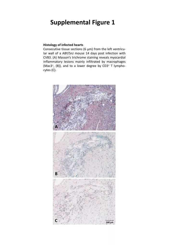
Supplemental Figure 1. Histology of infected hearts

E N D
Supplemental Figure 1 Histology of infected hearts Consecutive tissue sections (6 µm) from the left ventricu-lar wall of a ABY/SnJ mouse 14 days post infection with CVB3. (A) Masson’s trichrome staining reveals myocardial inflammatory lesions mainly infiltrated by macrophages (Mac3+, (B)), and to a lower degree by CD3+T lympho-cytes (C). A B C 100 mm