Download
1 / 24
250 likes | 411 Views
By Jane Horlings. Microscopy. Microscopy. Robert Hooke, circa 1700s. Electron Microscope, late 1900s. Light Microscopes. Early light microscope (UL), drawing by Hooke (LL). Light Microscopes. Dissecting (stereo) microscope (L), compound microscope (R). Light Microscopy; Plant Cells.

E N D
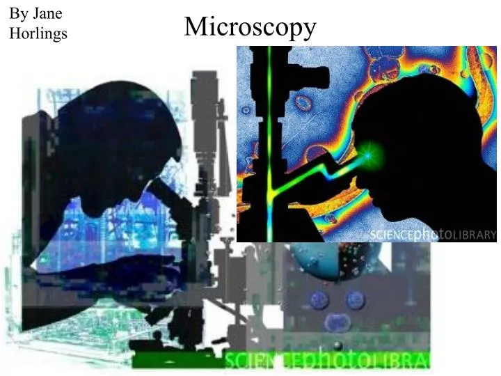
By Jane Horlings Microscopy
Microscopy Robert Hooke, circa 1700s Electron Microscope, late 1900s
Light Microscopes Early light microscope (UL), drawing by Hooke (LL)
Light Microscopes Dissecting (stereo) microscope (L), compound microscope (R)
Light Microscopy; Plant Cells Onion root tip, cell division (L), shoot tip (R)
Light Microscopy, Phase Contrast Single celled Amoeba dividing (L), green alga Micrasterias (R)
Light Microscopy Fluorescence Confocal Phase contrast
Limitations of Electron Microscopy • Works in a vacuum • Specimens are dead, chemically preserved; no life processes can be seen • No color (colorized by artist on computer)
Electron Microscopes Transmission Electron Microscope (TEM) Scanning Electron Microscope (SEM)
Scanning Electron Microscopy TEM of cilia SEM of cilia
Bacterium SEM RBCs in clot
SEM Mite (UL) Gecko toes (LL) Shark skin (LR)
SEM Trypanosome Red blood cells Red blood cells and Trypanosoma (L), Giardia (R)
Transmission Electron Microscopy TEM of cilia SEM of cilia
TEM Viruses (L), animal cell (R)
TEM Animal cell (L), muscle tissue (R)
Light Microscopy • Based on light • Specimens can be alive; life processes can be seen • Color; dyes may be used
Parts of the Microscope • Ocular lenses • Objective lenses • How to compute the magnification
Use of the Microscope • Place slide in center • Adjust light, lenses, barrel • Put on low magnification! • Move stage all the way up and then back down half a turn!
Use of the Microscope • Look and readjust focus • Move to higher magnification if needed
Use of the Microscope • Importance of focusing with the fine adjustment! • Oil immersion lens • How to adjust the light