Download
1 / 28
280 likes | 300 Views
Explore the structure and function of the heart, arteries, veins, and capillaries in the circulatory system. Learn about blood flow, cardiac conduction, regulation of the cardiac cycle, heart sounds, and hemodynamics.

E N D
The Cardiovascular System Kimberly Ball,Keisha Blanco, & Joshua Hyun Period 3
Introduction of the Circulatory System • System made up of the heart and different vessels • Heart • Arteries • Capillaries • Veins • Transports blood • Carries nutrients and oxygen • Maintains Homeostasis • Allows for function of organs
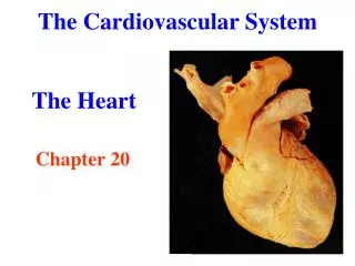
Size and Location • Heart • Size: • Individual’s fist closed • 14 centimeters • Location: • Chest Cavity between lungs • Left of the center • Base is on the wide superior border • Apex is found at the inferior point
Structure of The Heart • Heart: surrounded by serous membrane • Three Serous membranes of tissue • Visceral Pericardium • Pericardial Cavity • Parietal • Fibrous Tissue • Heart Wall 3 layers • Epicardium • Myocardium • Endocardium
Structure of The Heart Cont. • Chambers of the Heart • 2 Atria Upper Chambers • Left Atrium • Right Atrium • 2 Ventricles Lower Chambers • Left Ventricle • Right Ventricle • Heart: 4 Great Vessels • Superior Vena Cava • Inferior Vena Cava • Pulmonary Trunk • Aorta Pump
Structure of The Heart Cont. • Heart: Major Blood Vessels • Arteries • Aorta • Pulmonary arteries • Coronary Arteries • Veins • Superior and Inferior Vena Cava • Coronary Sinus • Pulmonary Veins
Structure of The Heart Cont. • Heart: Valves • Atrioventricular • Tricuspid • Bicuspid • Chordae Tendineae • Papillary Muscle • Semilunar Valves • Pulmonary SL Valve • Aortic SL Valve
Blood Flow • The Blood follows a certain path. • Vena Cava (Deoxygenated Blood) • Right Atrium (Deoxygenated Blood) • Right Ventricle (Deoxygenated Blood) • Pulmonary Artery (Deoxygenated Blood) • Lungs (Mixed Blood) • Pulmonary Vein (Oxygenated Blood) • Left Atrium (Oxygenated Blood) • Left Ventricle (Oxygenated Blood) • Aorta (Oxygenated Blood) • Blood Capillaries (Mixed Blood)
Bloodflow cont. • Pulmonary and Coronary • Pulmonary circuit • -system of blood vessels • that carries blood between • the heart and the lungs • Coronary circuit • -pathway in which the • the heart muscle is supplied • with blood
Cardiac Conduction System • Autorhythmic= self exciting • Cells that make-up CCS are in charge of initiating and distributing cardiac impulses throughout the heart muscle= heart to beat • Sinoatrial Node • Atrioventricular Node • Atrioventricular Bundle • Right and Left Bundle Branches • Purkinje Fibers
Electrocardiogram (ECG) -The electrical changes that happen in the myocardium during a cycle of cardiac are recorded in the electrogastrogram.
CARDIAC CYCLE • What is the Cardiac Cycle? • -Events that lead to complete a full complete heartbeat is known as a heartbeat. • Systole is a phase for contraction • Diastole is an action for relaxation • The Cardiac Cycle has Phases • Relaxation Period • Ventricular Filling • Ventricular Systole
Heart Sounds • stethoscope “lubb-dupp” • Types of Sounds: • lub= closed AV valves • dub= closed SL valves • murmur= incomplete closing valves-> blood leakage Normal Heart Sound Visualization https://www.youtube.com/watch?v=dDg7GDpR1RE
Regulation of Cardiac Cycle & the Heart Rate A. Autonomic Nervous System: 1. parasympathetic 2. sympathetic B. Chemicals 1. hormones tend to increase 2. ions a. calcium tends to increase b. both potassium and sodium decrease C. Age (decreases) D. Sex 1. females= increases 2. males= decreases E. Temperature F. Emotion G. Disease
Types of Blood Vessels • Arteries • 3 Layers: • 1) tunica interna (intima) • 2) tunica media • 3) tunica externa (adventitia) • Arterioles • Regulates Blood Pressure: • A) Vasoconstruction (contraction) • B) Vasodilation • Capillaries • 3 Types: • 1)Continuous Capillary • 2)Fenestrated Capillary • 3)Sinusoids
Type of Blood Vessels Cont. • Veins • - 3 thin walled vessels • 1) tunica intima • 2)tunica media • 3)tunica externa • -Valves • Prevent backflow
Hemodynamics • A. Blood Pressure • taken out by blood on the wall of blood vessel. • maximum pointduringsystole(contraction) • lowestpoint duringdiastole (relaxation). • MABP = 120 mm Hg/ 80 mm Hg B. Influence Arterial Blood Pressure 1. Heart Action-> cardiac output • Cardiac Output is affected by: • * stroke volume (SV)/ heart rate (HR) 2.. Peripheral Resistance is the opposition to blood flow • Depends on three things: · Blood viscosity · Total blood vessel length · Blood Vessel Radius
Hemodynamics Cont. C. Regulation of Blood Pressure and Blood Flow: 1. Neural Regulation: 2. Hormonal Control Hormones that increase BP: ·Epinephrine ·norepinephrine ·Angiotensin II ·Aldosterone D. Checking Circulation: 1. Pulse b. found at wrist c. Normal pulse-> between 70-80 bpm Hormones that decrease BP: ·Atrial natriuretic peptide (ANP) ·Histamine
Paths of Circulation • A. Pulmonary Circuit 1. pulmonary trunk 2. right and left pulmonary arteries 3. capillaries in lungs 4. right and left pulmonary veins • B. Systemic Circuit • 1. Arterial System • Venous System • - Superior Vena Cava • - Inferior Vena Cava
FYI: Veins are parallel to the arteries FYI: Veins are parallel to the arteries
Life Span Changes • When one becomes older • cholesterol begins to seep into the arteries • cardiac cells are exchanged for fibrous connective tissue and fat. • resting heart rate decreases while the blood pressure increases.
Works Cited Heart Care Centre. Kenning Corporation, 9 Jan. 2007. Web. 04 Mar. 2015. <http://www.heartcarecentre.co.nz/core/node/19>. Heart Muscle. University of Illinois in Chicago, 17 May 2009. Web. 04 Mar. 2015. <https://www.uic.edu/classes/phyb/phyb516/BaranyUpdate4/Heart/Heart.html>. Ivy Rose Holistic. Ivy Rose, 9 Nov. 2003. Web. 4 Mar. 2015. <http%3A%2F%2Fwww.ivyroses.com%2FHumanBody%2FBlood%2FHeart_Structure.php>. National Career Institute. National Institutes of Health, 13 Nov. 2003. Web. 4 Mar. 2015. <http%3A%2F%2Ftraining.seer.cancer.gov%2Fanatomy%2Fcardiovascular%2Fheart%2Fstructure.html>. On X Life Technologies Inc. The American Heart Association, 23 Aug. 2005. Web. 4 Mar. 2015. <http%3A%2F%2Fwww.onxlti.com%2Fpatient-guide%2Fblood-flow-heart%2F>. Practice Learning Resources. The University of Nottingham, 5 Dec. 2007. Web. 04 Mar. 2015. <http://www.nottingham.ac.uk/NURSING/practice/resources/cardiology/function/conduction.php>.