Download
1 / 54
540 likes | 573 Views
Explore the classification of joints, from fibrous to synovial, and learn how they facilitate movement, with examples and special features explained in detail. Discover the difference between sprains and strains, and the specific characteristics of various joint types such as planar, hinge, pivot, and condyloid joints. Get insight into common joint injuries and treatments like arthroscopy and arthroplasty in this comprehensive guide to joint anatomy and function.

E N D
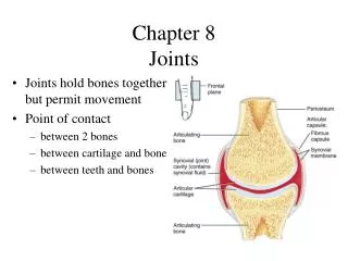
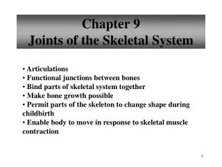
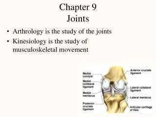
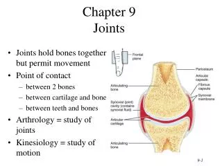
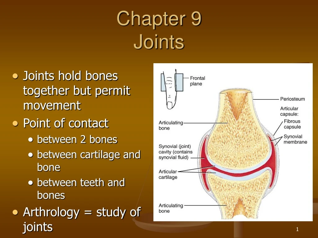
Chapter 9Joints • Joints hold bones together but permit movement • Point of contact • between 2 bones • between cartilage and bone • between teeth and bones • Arthrology = study of joints • Kinesiology = study of motion
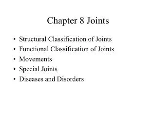
Classification of Joints • Structural classification based upon: • presence of space between bones • type of connective tissue holding bones together • collagen fibers • cartilage • joint capsule & accessory ligaments • Functional classification based upon movement: • immovable = synarthrosis • slightly movable = amphiarthrosis • freely movable = diarthrosis
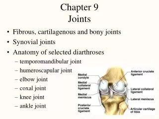
Fibrous Joints • Lack a synovial cavity • Bones held closely together by fibrous connective tissue • Little or no movement (synarthroses or amphiarthroses) • structural type • sutures • syndesmoses • gomphoses
Sutures • Thin layer of dense fibrous connective tissue unites bones of the skull • Immovable (synarthrosis) • If fuse completely in adults is synostosis
Syndesmosis • Fibrous joint • bones united by ligament • Slightly movable (amphiarthrosis) • Anterior tibiofibular joint and Interosseous membrane
Gomphosis • Ligament holds cone-shaped peg in bony socket • Immovable (amphiarthrosis) • Teeth in alveolar processes
Cartilaginous Joints • Lacks a synovial cavity • Allows little or no movement • Bones tightly connected by fibrocartilage or hyaline cartilage • 2 types • synchondroses • symphyses
Synchondrosis • Connecting material is hyaline cartilage • Immovable (synarthrosis) • Epiphyseal plate or joints between ribs and sternum
Symphysis • Fibrocartilage is connecting material • Slightly movable (amphiarthroses) • Intervertebral discs and pubic symphysis
Synovial Joints • Synovial cavity separates articulating bones • Freely moveable (diarthroses) • Articular cartilage • reduces friction • absorbs shock • Articular capsule • surrounds joint • thickenings in fibrouscapsule called ligaments • Synovial membrane • inner lining of capsule • secretes synovial fluid containing hyaluronic acid slippery) • brings nutrients to articular cartilage
Example of Synovial Joint • Joint space is synovial joint cavity • Articular cartilage covering ends of bones • Articular capsule
Other Special Features • Accessory ligaments • extracapsular ligaments • outside joint capsule • intracapsular ligaments • within capsule • Articular discs or menisci • attached around edges to capsule • allow 2 bones of different shape to fit tightly • increase stability of knee - torn cartilage • Bursae = saclike structures between structures • skin/bone or tendon/bone or ligament/bone
Arthroscopy & Arthroplasty • Arthroscopy = examination of joint • instrument size of pencil • remove torn knee cartilages & repair ligaments • small incision only • Arthroplasty = replacement of joints • total hip replaces acetabulum & head of femur • plastic socket & metal head • knee replacement common
Torn Cartilage and Arthroscopy • Damage to menisci of the knee joint • Visualization of the inside of a joint • arthroscope • requires only small incisions • Repair may include removal of torn cartilage
Nerve and Blood Supply • Nerves to joints are branches of nerves to nearby muscles • Joint capsule and ligaments contain pain fibers and sensory receptors • Blood supply to the structures of a joint are branches from nearby structures • supply nutrients to all joint tissues except the articular cartilage which is supplied from the synovial fluid
Sprain versus Strain • Sprain • twisting of joint that stretches or tears ligaments • no dislocation of the bones • may damage nearby blood vessels, muscles or tendons • swelling & hemorrhage from blood vessels • ankle if frequently sprained • Strain • less serious injury • overstretched or partially torn muscle
Planar Joint • Bone surfaces are flat or slightly curved • Side to side movement only • Rotation prevented by ligaments • Examples • intercarpal or intertarsal joints • sternoclavicular joint • vertebrocostal joints
Hinge Joint • Convex surface of one bones fits into concave surface of 2nd bone • Uniaxial like a door hinge • Examples • Knee, elbow, ankle, interphalangeal joints • Movements produced • flexion = decreasing the joint angle • extension = increasing the angle • hyperextension = opening the joint beyond the anatomical position
Pivot Joint • Rounded surface of bone articulates with ring formed by 2nd bone & ligament • Monoaxial since it allows only rotation around longitudinal axis • Examples • Proximal radioulnar joint • supination • pronation • Atlanto-axial joint • turning head side to side “no”
Condyloid or Ellipsoidal Joint • Oval-shaped projection fits into oval depression • Biaxial = flex/extend or abduct/adduct is possible • Examples • wrist and metacarpophalangeal joints for digits 2 to 5
Abduction and Adduction Condyloid joints Ball and Socket joints
Saddle Joint • One bone saddled-shaped; other bone fits as a person would sitting in that saddle • Biaxial • Circumduction allows tip of thumb travel in circle • Opposition allows tip of thumb to touch tip of other fingers • Example • trapezium of carpus and metacarpal of the thumb
Ball and Socket Joint • Ball fitting into a cuplike depression • Multiaxial • flexion/extension • abduction/adduction • rotation • Examples • shoulder joint • hip joint
Bursae and Tendon Sheaths • Bursae • fluid-filled saclike extensions of the joint capsule • reduce friction between moving structures • skin rubs over bone • tendon rubs over bone • Tendon sheaths • tubelike bursae that wrap around tendons at wrist and ankle where many tendons come together in a confined space • Bursitis • chronic inflammation of a bursa
Summary of Movements at Synovial Joints • Gliding • no change in angle of joint • Angular movements • increase or decrease in angle between articulating bones • flexion, extension, hyperextension • adduction, abduction • circumduction is a combination of above movements • Rotation • bone revolves around its own axis • Special movements • uniquely named movements for jaw, hand and foot
Circumduction • Movement of a distal end of a body part in a circle • Combination of flexion, extension, adduction and abduction • Occurs at ball and socket, saddle and condyloid joints
Rotation • Bone revolves around its own longitudinal axis • medial rotation is turning of anterior surface in towards the midline • lateral rotation is turning of anterior surface away from the midline • At ball & socket and pivot type joints
Special Movements of Mandible • Elevation = upward • Depression = downward • Protraction = forward • Retraction = backward
Special Hand & Foot Movements • Inversion • Eversion • Dorsiflexion • Plantarflexion • Pronation • Supination
Shoulder Joint • Head of humerus and glenoid cavity of scapula • Ball and socket • All types of movement
Glenohumeral (Shoulder) Joint • Articular capsule from glenoid cavity to anatomical neck • Glenoid labrum deepens socket • Many nearby bursa (subacromial)
Supporting Structures at Shoulder • Associated ligaments strengthen joint capsule • Transverse humeral ligament holds biceps tendon in place
Rotator Cuff Muscles • Attach humerus to scapula • Encircle the joint supporting the capsule • Hold head of humerus in socket
Elbow Joint • Hinge joint • trochlea notch of ulna and trochlea of humerus • flexion and extension of elbow • Pivot joint • head of radius and capitulum of humerus • supination and pronation of forearm
Articular Capsule of the Elbow Joint • Radial annular ligament hold head of radius in place • Collateral ligaments maintain integrity of joint
Hip Joint • Head of femur and acetabulum of hip bone • Ball and socket type of joint • All types of movement possible
Hip Joint Structures • Acetabular labrum • Ligament of the head of the femur • Articular capsule
Hip Joint Capsule • Dense, strong capsule reinforced by ligaments • iliofemoral ligament • ischiofemoral ligament • pubofemoral ligament • One of strongest structures in the body
Tibiofemoral Joint • Between femur, tibia and patella • Hinge joint between tibia and femur • Gliding joint between patella and femur • Flexion, extension, and slight rotation of tibia on femur when knee is flexed
Tibiofemoral Joint • Articular capsule • mostly ligs & tendons • Lateral & medial menisci = articular discs • Many bursa • Vulnerable joint • Knee injuries damage ligaments & tendons since bones do not fit together well
External Views of Knee Joint • Patella is part of joint capsule anteriorly • Rest of articular capsule is extracapsular ligaments • Fibular and tibial collateral ligaments
Intracapsular Structures of Knee • Medial meniscus • C-shaped fibrocartilage • Lateral meniscus • nearly circular • Posterior cruciate ligament • Anterior cruciate ligament
Temporomandibular Joint • Synovial joint • Articular disc • Gliding above disc • Hinge below disc • Movements • depression • elevation • protraction • retraction
Atlanto-occipital joints • Atlas and occipital condyles • Condyloid Joint • Flexion • Extension • Slight lateral tilting
Intervertebral Joints • Between bodies and intervertebral discs • symphysis • Between vertebral articular processes • synovial • Flexion • Extension • Lateral flexion
Elbow Joint • Trochlea of humerus, trochlear notch of ulna & head of radius • Pivot and hinge types • Flexion, extension, pronation & supination
Radiocarpal Joint • Articular disc • Condyloid type • Flexion, extension, abduction & adduction
Talocrural Joint • Tibia & fibula with talus • Hinge • Inversion, eversion, plantarflexion & dorsiflexion • Strong joint, seldom dislocates