Download
1 / 29
360 likes | 531 Views
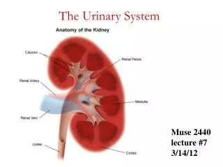
Histology of the Urinary System. Lecture Objectives. Describe the normal microscopic appearance of the different parts of the kidney including cortex, medulla, juxtaglomerular apparatus and the distribution of the vasculature within the kidney.

E N D
LectureObjectives • Describe the normal microscopic appearance of the different parts of the kidney including cortex, medulla, juxtaglomerular apparatus and the distribution of the vasculature within thekidney. • List the different parts of the nephron with the details of eachpart. • Describe the microscopical appearanceof the ureter, urinary bladder andurethra. • Compare the histological appearance ofthe distended and contractedbladder.
Internal Anatomy ofthe Kidneys • Parenchyma ofkidney • Renal cortex = superficial layer ofkidney • Renalmedulla • inner portion consisting of 8-18 cone-shaped renal pyramids separated by renal columns • renal papilla point toward center ofkidney • Medullary rays, parallel arrays of tubules penetrate the cortex from the base of the medullarypyramids • Drainage system fills renal sinuscavity • minor & major calyces empty into the renal pelvis which empties into theureter • minor calyces collect urine from the papillary ducts of thepapilla
Nephrons • A nephron consists of a renal corpuscle where fluid is filtered, and a renal tubule into which the filtered fluidpasses • Renal corpuscle = site of plasma filtration • glomerulus is capillaries where filtration occurs • glomerular (Bowman’s) capsule is double-walled epithelial cup that collects filtrate
Nephrons • A renal tubule consistsof: • proximal convoluted tubule (PCT) • loop of Henle (nephron loop) • distal convoluted tubule (DCT) • Distal convoluted tubules of several nephrons drain into to a single collecting duct and many collecting ducts drain into a small number of papillaryducts.
Nephrons • The loop ofHenle consistsof: • thick descendinglimb • thin descendinglimb • thin ascendinglimb • thick ascendinglimb
Nephrons:Types • There are two types of nephrons thathave differing structure andfunction • A corticalnephron • glomerulus in the outer portion of thecortex • short loop ofHenle • penetrates only into the outer region of themedulla • A juxtamedullarynephron • glomerulus deep in the cortex close to the medulla • long loop of Henle stretches through the medulla and almost reaches the renalpapilla
Cortical Nephron • 80-85% of nephrons are corticalnephrons • Renal corpuscles are in outer cortexand loops of Henle lie mainly incortex
Juxta- medullary Nephron • 15-20% of nephrons are juxtamedullarynephrons • Renal corpuscles close to medulla and long loops of Henle extend into deepest medulla enabling excretion of dilute or concentratedurine
RenalCorpuscle • Renal Corpuscle is about 200 μm indiameter • Consistsof: • Bowman’s capsule surrounds capsular (urinary)space • podocytes cover capillaries to form viscerallayer • simple squamous cells form parietal layer ofcapsule • Glomerular capillaries arise from afferent arteriole & form a ball before emptying into efferentarteriole • Fenestrated endothelial cells constitute thecapillaries • Renal corpuscle have a vascular pole where the afferent arteriole enters and the efferent arteriole leaves, and a urinary pole where proximal convoluted tubulebegins
GlomerularCapsule • The glomerular capsule consists of visceral and parietallayers • The parietal layer consists of simple squamous epithelium supported by basal lamina and thin layer of reticular fibers • Near the urinary pole the epithelium becomes cuboidal or lowcolumnar • The visceral layer consists of modified simple squamous epithelial cells called podocytes.
GlomerularCapsule • Podocytes have cell body from which arise several primary processes • Each primary process gives rise to many secondary processes (pedicels) • Only the secondary processes have direct contact with the basal lamina • Filtration slits , is a 25 nm wide elongated spaces between the secondaryprocesses • Filtration slits are covered by 6 nm thickdiaphragm • A thick basement membrane (0.1 μm) separates the capillaries endothelium and thepodocytes • Lamina densa covered by lamina rara on eachside • Basement membrane is selectively permeable formacromolecules
GlomerularCapillaries • Glomerular capillaries have mesangial cells adhere to theirwalls • mesangial cells are contractile cells and have receptors for angiotensin II, so it can reduce the glomerularflow • mesangial cells contains also receptors for natriuretic factor produced by the cardiac atria cells causing vasodilation and relaxes mesangialcells • mesangial cells also have severalfunctions • Support the glomerulus • Produce extracellularmatrix • Immune response • Outside the glomerulus in the vascular pole, form the extraglomerular mesangial cells, which is part of the extraglomerularapparatus
Proximal convolutedtubules • Longer than the distal convoluted tubule, thus seen more near the renal corpuscle in thecortex • Consists of cuboidalcells • Acidophilic cytoplasm , numerous elongated mitochondria • Brush border, abundant microvilli • Large cells, 3-5 surround each tubule • Lateral interdigitation connecting lateral walls of cells, difficult to distinguish boundaries between adjacent cells • Basal infoldings with mitochondria (basal striations), characteristic of cells with active ionictransport
Proximal convolutedtubules • Functions • Resorption of 80% water, electrolytes through Na+, Ka+- ATPase • Resorption of 100% glucose and aminoacids • Protein resorption: apical canaliculi connecting microvilli increase the absorption of macromolecules by pinocytosis. • Vacuoles and lysosomes are more abundant in the apical portion of the cytoplasm • Secretion of creatinine and organic acids and bases, so pH modification
Loop ofHenle • Thick descending limb (60μm) • Cytology • Similarto proximal convolutedtubules • Slightly fewer microvilli • Less basolateralinterdigitation • Mitochondria are smaller and more randomly oriented • Function: Na+ and H2Oreabsorption • Thin tubule (12μm) • Cytology: Simple squamousepithelium • Function • Descending limb: permeable to H2O andsalt • Ascending limb: impermeable to H2O, permeable to salt andurea • Concentration of theurine
Loop ofHenle • Thick ascending limb • Cytology • Cuboidal cells with apicalnuclei • Numerous basal infoldings withlarger mitochondria • Few and shortmicrovilli • Macula densa, part ofjuxtaglomerular apparatus • Function • Cl- and Na+ transport into basolateral spaces • Establish a salt concentration gradientin medulla for urineconcentration
Distal convoluted tubule • Cytology • Same as thickascending limb of loop ofHenle • Cells are flatter and smaller than those of proximal convoluted tubules, so more cells will appear in cross section • Function • Na+ reabsorption, Ka+ secretion(aldosterone) • Reabsorption of bicarbonate, hydrogen secretion, pH increases • Conversion of ammonia to ammoniumions
Collecting tubulesand ducts • Types • Connecting tubule: 40 µm in diameter, in cortex • Cortical duct: medullaryrays • Medullary duct:medulla • Papillary duct (of Bellini): 200 µm in diameter, apex • Epithelium: cuboidal to columnarcells • Principal cells: light stained cells, cilium, short microvilli, basal infoldings, permeable to H2O (H2O channel, in the presence of antidiuretic hormone) • Intercalated cells: dark stained cells, microplicae (folds), H+ secretion, bicarbonate reabsorption. Absent in inner medulla
JuxtaglomerularApparatus • Senses and regulates blood flow andcomposition • Structure where afferent arteriole makes contact with ascending limb of loop ofHenle • Consists of three celltypes • juxtaglomerular cells are modified muscle cells in arteriole, renin secretion • macula densa: sense NaCl concentration, regulates reninrelease. • It is thickened part of ascendinglimb • the extraglomerular mesangial cells: unknownfunction
Regulation of blood flow by renin Stimulation → JG cells → Renin→ • Angiotensinogen → Angiotensin I (lung)→ Angiotensin II → Aldosterone (Adrenal cortex) → Na+, Cl- uptake(DCT)
Blood & Nerve Supply ofKidney • Abundantly supplied with bloodvessels • receive 25% of resting cardiac output via renalarteries • Functions of different capillarybeds • glomerular capillaries where filtration of bloodoccurs • vasoconstriction & vasodilation of afferent & efferent arterioles produce large changes in renalfiltration • peritubular capillaries that carry away reabsorbed substances fromfiltrate • vasa recta supplies nutrients to medulla without disrupting its osmolarityform • The nerve supply to the kidney is derived from the renal plexus (sympathetic division of ANS). Sympathetic vasomotor nerves regulate blood flow & renal resistance by alteringarterioles
Urinary bladder & Urinarypassages • Bladder and urinary passages store and conduct urine to the exterior • All have the same histologicalstructure: • Themucosa • Transitionalepithelium • Lamina propria • Dense woven sheath of smoothmuscle • Calyces, renal pelvis and ureters: helicalarrangement • Distal part of ureter:longitudinal • Bladder: run in every direction • Bladderneck: • Internal longitudinal. Distal to bladder neck becomecircular • Surround prostaticurethra • Extend to external urethral meatus inwomen • Middle circular: ends at the bladderneck • Outer longitudinal layer: continue to the prostate in men and to the external urethral meatus inwomen • Adventitia • Serosa: upper part of thebladder
Emptybladder Fullbladder ureter
Urethra • Male urethra: consists of fourparts • Prostatic urethra • Close to bladder, and ducts of prostate gland opens into it • Transitionalepithelium • Dorsal and distal part have verumontanum (elevation protrudes into its interior) • – Closed tube (prostatic utricle) opens into the tip with no knownfunction • Ejaculatory ducts open on the sides of theverumontanum • Membranous urethra • 1 cm, surrounded by the external urethra sphincter (striatedmuscle) • Stratified or pseudostratified columnarepithelium • Bulbous and pendulous parts ofurethra • Located in the corpus spongiosum of thepenis • Lumen dilates distally forming the fossanavicularis • Pseudostratified columnar with stratified squamousareas • Litter’s glands: mucous glands found along the entire urethra and mostly in the pendulouspart • Femaleurethra • 4-5 cm long tube, lined by stratified squamous epithelium and areas of pseudostratified columnarepithelium • The mid part is surrounded by external urethralsphincter