Download
1 / 36
470 likes | 2.13k Views
Understanding Coral Histology - How & Why -. Ha-Rim Cha Division of Invertebrate Zoology The Natural History Museum of University of Kansas and Biodiversity Research Center. Coral Slide Reading Workshop. To learn histological techniques & slide reading skills for scleractinians.

E N D
Understanding Coral Histology- How & Why - Ha-Rim Cha Division of Invertebrate Zoology The Natural History Museum of University of Kansas and Biodiversity Research Center
Coral Slide Reading Workshop • To learn histological techniques & slide reading skills for scleractinians • Mote Marine Laboratory, Summerland Key, FL
Cylindrical body Family Sideractiidae Family Corallimorphidae Plate-like body Family Ricordeidae Family Discosomatidae Phylogeny of Corallimorpharia • Questions about relationships within the order Vincent B. Hargreaves Vincent B. Hargreaves
Questions about relationships between Corallimorpharia and two closest orders Actiniaria (sea anemones) and Scleractinia (stony corals) Phylogeny of Corallimorpharia ↑Stony coral Corallimorpharia↑ ↑ Sea anemones
Characters to be examined • Calcareous exoskeleton • Polyp shape / size • Tentacle shape / number / arrangement • Type of mesenteries • Mesentery number / arrangement • Development of musculature • Nematocyst composition • Life style – solitary or colonial • Habitat • Endosymbiotic relationships Scott R. Santos
Characters known about Coral Anatomy • Calcareous exoskeleton • Polyp shape / size • Tentacle shape / number / arrangement • Type of mesenteries • Mesentery number / arrangement • Development of musculature • Nematocyst composition • Life style – solitary or colonial • Habitat • Endosymbiotic relationships
Characters used for Coral Taxonomy & Phylogeny • Taxonomy of scleractinian corals is based on their skeletal structure Copyright @ Brin Edwards
Characters used for Coral Taxonomy & Phylogeny • Taxonomy of scleractinian corals Genus Fungia Genus Tubastrea
Characters used for Coral Taxonomy & Phylogeny • Order level phylogeny of Hexacorallia Scleractinia ? Corallimorpharia Actiniaria
Phylogeny • Order level phylogeny of Hexacorallia a. Chen et al. (1995): 28S rRNA b. Fautin & Lowenstein (1994): radioimmunoassay c. France et al. (1996): 16S rRNA d. Daly et al. (2003): Morphology, 16S, 18S and 28S
Microscopic Polyp Anatomy • Additional characters on coral morphology • An independent character set to compare with published molecular data
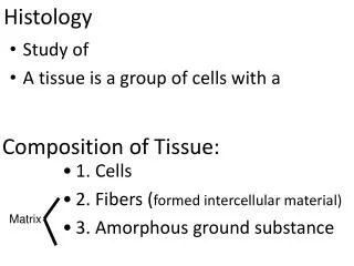
Histology • Histology (= Microscopic anatomy) is… the study of cells, tissues, organs, and organ systems of plants and animals
Histology • Histology (= Microscopic anatomy) is… the study of cells, tissues, organs, and organ systems of plants and animals Structure & Composition of an organism or a part reflects its key functions (e.g. respiration, metabolism, reproduction, movement)
Coral Histology • To support studies on ecology, physiology, reproduction, biochemistry, immunology, embryology, and systematics • Histopathology -- Study on coral diseases (e.g. white-band disease, black-band disease, red-band disease) or bleaching event
Bleaching & Coral Disease Bleaching Black-Band Disease DEAD ALIVE! Yellow-Band Disease ( photos by E. Peters)
Procedure for coral histology Staining Collecting Sectioning Fixation Decalcifying and Trimming Embedding
Specimen Collection • Using a cold chisel and a coring device (for a large colony) • Relax the collected specimens • Obtaining permission prior to conducting the study • Reporting export and import of collected materials to the United States Fish and Wildlife Service is required
Fixation • To preserve the structure and composition of the tissue • Post-mortem effects are usually prevented by denaturing and precipitating protein, or by cross-linking the proteins with aldehyde and/or other chemicals
Fixation • The choice of fixative will depend on the embedding medium to be used. • for light microscopy, paraffin embedding: Modified Helly’s solution, Bouin’s solution, and 10% seawater Formalin • for electron microscopy only: Glutaraldehyde/Paraformaldehyde Solution
Decalcifying & Trimming • Embedding specimens prior to decalcification using 1~1.5% low-melting point agarose or HistoGel (Optional) • Decalcifying solutionhydrochloric acid with a chelating agent, ethylene diamine tetraacetic acid (EDTA)
Processing for Embedding • Dehydrate to remove all traces of water • Infiltrate with a liquid that can be hardened sufficiently to allow cutting of thin section. • Clear with a reagent that is miscible with the dehydrating solution and the embedding medium
Embedding • Embedding medium: Paraffin for light microscopy & Glycol methacrylate or epoxy for electron microscopy • Topology of tissue (Humason, 1967)
Sectioning • Cut sections at 6-10 micrometers or thinner • Remove wrinkles and air bubbles from the ribbon by floating on warm-water • A set of sections are picked up on the slide and dried (Humason, 1967)
Staining • To visualize cell and tissue composition and structure • Stains are used to distinguish the various cell and tissue components; they bind preferentially depending on the biochemistry of the organelles, membranes, etc.
Staining • “routine” stain 1) Hematoxylin & Eosin2) Heidenhain’s Azocarmine-Aniline Blue3) Movat’s Pentachrome • Special stains are used to identify - particular components or microorganisms- heavy metal impregnation- immunohistochemistry or autoradiography
Hematoxylin & Eosin staining Actinodiscus fungiformis
Staining • “routine” stain 1) Hematoxylin & Eosin2) Heidenhain’s Azocarmine-Aniline Blue3) Movat’s Pentachrome • Special stains are used to identify - particular components or microorganisms- heavy metal impregnation- immunohistochemistry or autoradiography
Heidenhain’s staining Genus Megalactis (Photo by Dr. Adorian Ardelean)
Staining • “routine” stain 1) Hematoxylin & Eosin2) Heidenhain’s Azocarmine-Aniline Blue3) Movat’s Pentachrome • Special stains are used to identify - particular components or microorganisms- heavy metal impregnation- immunohistochemistry or autoradiography
Movat’s Pentachrome staining Astrangia poculata (photo by Dr. Esther Peters)
Staining • “routine” stain 1) Hematoxylin & Eosin2) Heidenhain’s Azocarmine-Aniline Blue3) Movat’s Pentachrome • Special stains are used to identify - particular components or microorganisms- heavy metal impregnation- immunohistochemistry or autoradiography
Coral Slide Reading • The 2-dimensional images seen in light microscopy came from 3-dimensional structures • Interpreting the appearance require careful consideration of several factors: what is known about the specimens, the normal appearance of the cells and tissues, and changes that might have affected their conditions.
Coral Slide Reading • Details and variation of internal anatomy • Reproductive pattern • Symbiotic relationships • Biomechanics • Health of corals Get the CHARACTERS to reconstruct the phylogeny!
Actiniaria, Corallimorpharia, Scleractinia Actiniaria Genus Megalactis (Photo by Dr. Adorian Ardelean) (photo by Dr. Esther Peters) Corallimorpharia Corynactis californica Scleractinia Astrangia poculata
Scleractinian corals Genus Oculina Genus Pocillipora ADD MOREPICTURES ADD MORE KNOWLEDGE ADD MORE CHARACTERS BETTER CLASSIFICATION & PHYLOGENY
Acknowledgements • Panorama Small Grant, KUNHM & BRC • Graduate Travel Fund, EEB, KU • NSF grant 9978106 in the program Partnerships to Enhance Expertise in Taxonomy (PEET) to Dr. Daphne Fautin • Dr. Esther Peters, Tetra Tech Inc. • Dr. Daphne Fautin, Dr. Meg Daly and Dr. Adorian Ardelean Vincent B. Hargreaves Adorian Ardelean George Miller