Download
1 / 64
1.03k likes | 2.77k Views
Chapter 21 The Cardiovascular System: Blood Vessels and Hemodynamics. Structure and function of blood vessels Hemodynamics forces involved in circulating blood Major circulatory routes. Anatomy of Blood Vessels. Closed system of tubes that carries blood

E N D
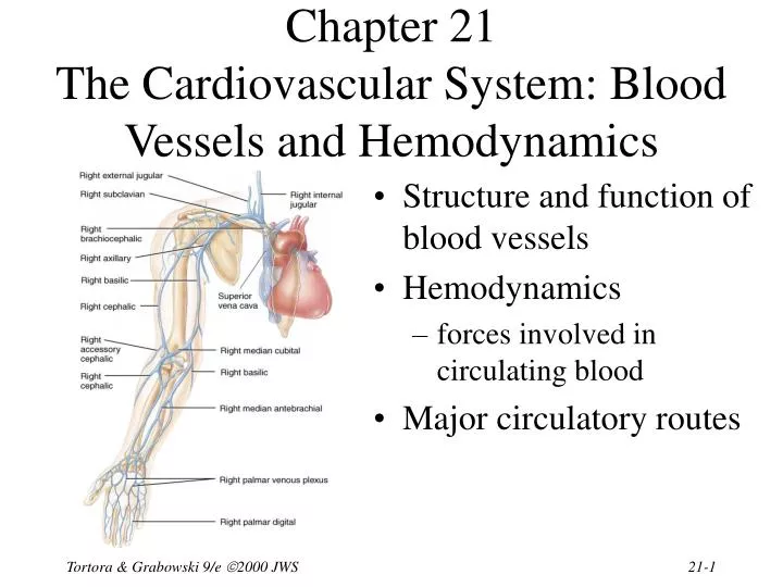
Chapter 21The Cardiovascular System: Blood Vessels and Hemodynamics • Structure and function of blood vessels • Hemodynamics • forces involved in circulating blood • Major circulatory routes Tortora & Grabowski 9/e 2000 JWS
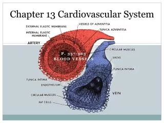
Anatomy of Blood Vessels • Closed system of tubes that carries blood • Arteries carry blood from heart to tissues • elastic arteries • muscular arteries • arterioles • Capillaries are thin enough to allow exchange • Venules merge to form veins that bring blood back to the heart • Vasa vasorum is vessels in walls of large vessel Tortora & Grabowski 9/e 2000 JWS
Arteries • Tunica interna (intima) • simple squamous epithelium known as endothelium • basement membrane • internal elastic lamina • Tunica media • circular smooth muscle & elastic fibers • Tunica externa • elastic & collagen fibers Tortora & Grabowski 9/e 2000 JWS
Sympathetic Innervation • Vascular smooth muscle is innervated by sympathetic nervous system • increase in stimulation causes muscle contraction or vasoconstriction • decreases diameter of vessel • injury to artery or arteriole causes muscle contraction reducing blood loss (vasospasm) • decrease in stimulation or presence of certain chemicals causes vasodilation • increases diameter of vessel • nitric oxide, K+, H+ and lactic acid cause vasodilation Tortora & Grabowski 9/e 2000 JWS
Elastic Arteries • Largest-diameter arteries have lot of elastic fibers in tunica media • Help propel blood onward despite ventricular relaxation (stretch and recoil -- pressure reservoir) Tortora & Grabowski 9/e 2000 JWS
Muscular Arteries • Medium-sized arteries with more muscle than elastic fibers in tunica media • Capable of greater vasoconstriction and vasodilation to adjust rate of flow • walls are relatively thick • called distributing arteries because they direct blood flow Tortora & Grabowski 9/e 2000 JWS
Arterioles • Small arteries delivering blood to capillaries • tunica media containing few layers of muscle • Metarterioles form branches into capillary bed • to bypass capillary bed, precapillary sphincters close & blood flows out of bed in thoroughfare channel • vasomotion is intermittent contraction & relaxation of sphincters that allow filling of capillary bed 5-10 times/minute Tortora & Grabowski 9/e 2000 JWS
Capillaries form Microcirculation • Microscopic vessels that connect arterioles to venules • Found near every cell in the body but more extensive in highly active tissue (muscles, liver, kidneys & brain) • entire capillary bed fills with blood when tissue is active • lacking in epithelia, cornea and lens of eye & cartilage • Function is exchange of nutrients & wastes between blood and tissue fluid • Structure is single layer of simple squamous epithelium and its basement membrane Tortora & Grabowski 9/e 2000 JWS
Types of Capillaries • Continuous capillaries • intercellular clefts are gaps between neighboring cells • skeletal & smooth, connective tissue and lungs • Fenestrated capillaries • plasma membranes have many holes • kidneys, small intestine, choroid plexuses, ciliary process & endocrine glands • Sinusoids • very large fenestrations • incomplete basement membrane • liver, bone marrow, spleen, anterior pituitary, & parathyroid gland Tortora & Grabowski 9/e 2000 JWS
Venules • Small veins collecting blood from capillaries • Tunica media contains only a few smooth muscle cells & scattered fibroblasts • very porous endothelium allows for escape of many phagocytic white blood cells • Venules that approach size of veins more closely resemble structure of vein Tortora & Grabowski 9/e 2000 JWS
Veins • Proportionally thinner walls than same diameter artery • tunica media less muscle • lack external & internalelastic lamina • Still adaptable to variationsin volume & pressure • Valves are thin folds of tunica interna designed to prevent backflow • Venous sinus has no muscle at all • coronary sinus or dural venous sinuses Tortora & Grabowski 9/e 2000 JWS
Varicose Veins • Twisted, dilated superficial veins • caused by leaky venous valves • congenital or mechanically stressed from prolonged standing or pregnancy • allow backflow and pooling of blood • extra pressure forces fluids into surrounding tissues • nearby tissue is inflamed and tender • Deeper veins not susceptible because of support of surrounding muscles Tortora & Grabowski 9/e 2000 JWS
Anastomoses • Union of 2 or more arteries supplying the same body region • blockage of only one pathway has no effect • circle of willis underneath brain • coronary circulation of heart • Alternate route of blood flow through an anastomosis is known as collateral circulation • can occur in veins and venules as well • Alternate routes to a region can also be supplied by nonanastomosing vessels Tortora & Grabowski 9/e 2000 JWS
Blood Distribution • 60% of blood volume at rest is in systemic veins and venules • function as blood reservoir • veins of skin & abdominalorgans • blood is diverted from it intimes of need • increased muscular activityproduces venoconstriction • hemorrhage causes venoconstriction to help maintain blood pressure • 15% of blood volume in arteries & arterioles Tortora & Grabowski 9/e 2000 JWS
Capillary Exchange • Movement of materials in & out of a capillary • diffusion (most important method) • substances move down concentration gradient • all plasma solutes except large proteins pass freely across • through lipid bilayer, fenestrations or intercellular clefts • blood brain barrier does not allow diffusion of water-soluble materials (nonfenestrated epithelium with tight junctions) • transcytosis • passage of material across endothelium in tiny vesicles by endocytosis and exocytosis • large, lipid-insoluble molecules such as insulin or maternal antibodies passing through placental circulation to fetus • bulk flow see next slide Tortora & Grabowski 9/e 2000 JWS
Bulk Flow: Filtration & Reabsorption • Movement of large amount of dissolved or suspended material in same direction • move in response to pressure • from area of high pressure to area of low • faster rate of movement than diffusion or osmosis • Most important for regulation of relative volumes of blood & interstitial fluid • filtration is movement of material into interstitial fluid • promoted by blood hydrostatic pressure & interstitial fluid osmotic pressure • reabsorption is movement from interstitial fluid into capillaries • promoted by blood colloid osmotic pressure • balance of these pressures is net filtration pressure Tortora & Grabowski 9/e 2000 JWS
Dynamics of Capillary Exchange 10 9 • Starling’s law of the capillaries is that the volume of fluid & solutes reabsorbed is almost as large as the volume filtered Tortora & Grabowski 9/e 2000 JWS
Net Filtration Pressure • Whether fluids leave or enter capillaries depends on net balance of pressures • net outward pressure of 10 mm Hg at arterial end of a capillary bed • net inward pressure of 9 mm Hg at venous end of a capillary bed • About 85% of the filtered fluid is returned to the capillary • escaping fluid and plasma proteins are collected by lymphatic capillaries (3 liters/day) Tortora & Grabowski 9/e 2000 JWS
Edema • An abnormal increase in interstitial fluid if filtration exceeds reabsorption • result of excess filtration • increased blood pressure (hypertension) • increased permeability of capillaries allows plasma proteins to escape • result of inadequate reabsorption • decreased concentration of plasma proteins lowers blood colloid osmotic pressure • inadequate synthesis or loss from liver disease, burns, malnutrition or kidney disease • Not noticeable until 30% above normal Tortora & Grabowski 9/e 2000 JWS
Hemodynamics • Factors affecting circulation • pressure differences that drive the blood flow • velocity of blood flow • volume of blood flow • blood pressure • resistance to flow • venous return • An interplay of forces result in blood flow Tortora & Grabowski 9/e 2000 JWS
Velocity of Blood Flow • Speed of blood flow in cm/sec is inversely related to cross-sectional area • blood flow is slower in thearterial branches • flow in aorta is 40 cm/sec whileflow in capillaries is .1 cm/sec • slow rate in capillaries allows forexchange • Blood flow becomes faster when vessels merge to form veins • Circulation time is time it takes a drop of blood to travel from right atrium back to right atrium Tortora & Grabowski 9/e 2000 JWS
Volume of Blood Flow • Cardiac output = stroke volume x heart rate • Other factors that influence cardiac output • blood pressure • resistance due to friction between blood cells and blood vessel walls • blood flows from areas of higher pressure to areas of lower pressure Tortora & Grabowski 9/e 2000 JWS
Blood Pressure • Pressure exerted by blood on walls of a vessel • caused by contraction of the ventricles • highest in aorta • 120 mm Hg during systole & 80during diastole • If heart rate increases cardiacoutput, BP rises • Pressure falls steadily insystemic circulation with distance from left ventricle • 35 mm Hg entering the capillaries • 0 mm Hg entering the right atrium • If decrease in blood volume is over 10%, BP drops • Water retention increases blood pressure Tortora & Grabowski 9/e 2000 JWS
Resistance • Friction between blood and the walls of vessels • average blood vessel radius • smaller vessels offer more resistance to blood flow • cause moment to moment fluctuations in pressure • blood viscosity (thickness) • ratio of red blood cells to plasma volume • increases in viscosity increase resistance • dehydration or polycythemia • total blood vessel length • the longer the vessel, the greater the resistance to flow • 200 miles of blood vessels for every pound of fat • obesity causes high blood pressure • Systemic vascular resistance is the total of above • arterioles control BP by changing diameter Tortora & Grabowski 9/e 2000 JWS
Factors that Increase Blood Pressure Tortora & Grabowski 9/e 2000 JWS
Venous Return • Volume of blood flowing back to the heart from the systemic veins • depends on pressure difference from venules (16 mm Hg) to right atrium (0 mm Hg) • tricuspid valve leaky andbuildup of blood on venousside of circulation • Skeletal muscle pump • contraction of muscles & presence of valves • Respiratory pump • decreased thoracic pressure and increased abdominal pressure during inhalation, moves blood into thoracic veins and the right atrium Tortora & Grabowski 9/e 2000 JWS
Syncope • Fainting or a sudden, temporary loss of consciousness not due to trauma • due to cerebral ischemia or lack of blood flow to the brain • Causes • vasodepressor syncope = sudden emotional stress • situational syncope = pressure stress of coughing, defecation, or urination • drug-induced syncope = antihypertensives, diuretics, vasodilators and tranquilizers • orthostatic hypotension = decrease in BP upon standing Tortora & Grabowski 9/e 2000 JWS
Control of Blood Pressure & Flow • Role of cardiovascular center • help regulate heart rate & stroke volume • specific neurons regulate blood vessel diameter Tortora & Grabowski 9/e 2000 JWS
Input to the Cardiovascular Center • Higher brain centers such as cerebral cortex, limbic system & hypothalamus • anticipation of competition • increase in body temperature • Proprioceptors • input during physical activity • Baroreceptors • changes in pressure within blood vessels • Chemoreceptors • monitor concentration of chemicals in the blood Tortora & Grabowski 9/e 2000 JWS
Output from the Cardiovascular Center • Heart • parasympathetic (vagus nerve) • decrease heart rate • sympathetic (cardiac accelerator nerves) • cause increase or decrease in contractility & rate • Blood vessels • sympathetic vasomotor nerves • continual stimulation to arterioles in skin & abdominal viscera producing vasoconstriction (vasomotor tone) • increased stimulation produces constriction & increased BP Tortora & Grabowski 9/e 2000 JWS
Neural Regulation of Blood Pressure • Baroreceptor reflexes • carotid sinus reflex • swellings in internal carotid artery wall • glossopharyngeal nerve to cardiovascular center in medulla • maintains normal BP in the brain • aortic reflex • receptors in wall of ascending aorta • vagus nerve to cardiovascular center • maintains general systemic BP • If feedback is decreased, CV center reduces parasympathetic & increases sympathetic stimulation of the heart Tortora & Grabowski 9/e 2000 JWS
Innervation of the Heart • Speed up the heart with sympathetic stimulation • Slow it down with parasympathetic stimulation (X) • Sensory information from baroreceptors (IX) Tortora & Grabowski 9/e 2000 JWS
Carotid Sinus Massage & Syncope • Stimulation (careful neck massage) over the carotid sinus to lower heart rate • paroxysmal superventricular tachycardia • tachycardia originating from the atria • Anything that puts pressure on carotid sinus • tight collar or hyperextension of the neck • may slow heart rate & cause carotid sinus syncope or fainting Tortora & Grabowski 9/e 2000 JWS
Chemoreceptor Reflexes • Carotid bodies and aortic bodies • detect changes in blood levels of O2, CO2, and H+ (hypoxia, hypercapnia or acidosis ) • causes stimulation of cardiovascular center • increases sympathetic stimulation to arterioles & veins • vasoconstriction and increase in blood pressure • Also changes breathing rates as well Tortora & Grabowski 9/e 2000 JWS
Hormonal Regulation of Blood Pressure • Renin-angiotensin-aldosterone system • decrease in BP or decreased blood flow to kidney • release of renin / results in formation angiotensin II • systemic vasoconstriction • causes release aldosterone (H2O & Na+ reabsorption) • Epinephrine & norepinephrine • increases heart rate & force of contraction • causes vasoconstriction in skin & abdominal organs • vasodilation in cardiac & skeletal muscle • ADH causes vasoconstriction • ANP (atrial natriuretic peptide) lowers BP • causes vasodilation & loss of salt and water in the urine Tortora & Grabowski 9/e 2000 JWS
Local Regulation of Blood Pressure • Local factors cause changes in each capillary bed • autoregulation is ability to make these changes as needed by demand for O2 & waste removal • important for tissues that have major increases in activity (brain, cardiac & skeletal muscle) • Local changes in response to physical changes • warming & decrease in vascular stretching promotes vasodilation • Vasoactive substances released from cells alter vessel diameter (K+, H+, lactic acid, nitric oxide) • systemic vessels dilate in response to low levels of O2 • pulmonary vessels constrict in response to low levels of O2 Tortora & Grabowski 9/e 2000 JWS
Shock and Homeostasis • Shock is failure of cardiovascular system to deliver enough O2 and nutrients • inadequate perfusion • cells forced to switch to anaerobic respiration • lactic acid builds up • cells and tissues become damaged & die Tortora & Grabowski 9/e 2000 JWS
Types of Shock • Hypovolemic shock due to loss of blood or body fluids (hemorrhage, sweating, diarrhea) • venous return to heart declines & output decreases • Cardiogenic shock caused by damage to pumping action of the heart (MI, ischemia, valve problems or arrhythmias) • Vascular shock causing drop inappropriate vasodilation -- anaphylatic shock, septic shock or neurogenic shock (head trauma) • Obstructive shock caused by blockage of circulation (pulmonary embolism) Tortora & Grabowski 9/e 2000 JWS
Homeostatic Responses to Shock • Mechanisms of compensation in shock attempt to return cardiac output & BP to normal • activation of renin-angiotensin-aldosterone • secretion of antidiuretic hormone • activation of sympathetic nervous system • release of local vasodilators • If blood volume drops by 10-20% or if BP does not rise sufficiently, perfusion may be inadequate -- cells start to die Tortora & Grabowski 9/e 2000 JWS
Restoring BP during Hypovolemic Shock Tortora & Grabowski 9/e 2000 JWS
Signs & Symptoms of Shock • Rapid resting heart rate (sympathetic stimulation) • Weak, rapid pulse due to reduced cardiac output & fast heart rate • Clammy, cool skin due to cutaneous vasoconstriction • Sweating -- sympathetic stimulation • Altered mental state due to cerebral ischemia • Reduced urine formation -- vasoconstriction to kidneys & increased aldosterone & antidiuretic hormone • Thirst -- loss of extracellular fluid • Acidosis -- buildup of lactic acid • Nausea -- impaired circulation to GI tract Tortora & Grabowski 9/e 2000 JWS
Evaluating Circulation • Pulse is a pressure wave • alternate expansion & recoil of elastic artery after each systole of the left ventricle • pulse rate is normally between 70-80 beats/min • tachycardia is rate over 100 beats/min/bradycardia under 60 • Measuring blood pressure with sphygmomanometer • Korotkoff sounds are heard while taking pressure • systolic blood pressure from ventricular contraction • diastolic blood pressure during ventricular contraction • provides information about systemic vascular resistance • pulse pressure is difference between systolic & diastolic • normal ratio is 3:2:1 -- systolic/diastolic/pulse pressure Tortora & Grabowski 9/e 2000 JWS
Pulse Points Tortora & Grabowski 9/e 2000 JWS
Systemic Circulation • All systemic arteries branch from the aorta • All systemic veins drain into the superior or inferior vena cava or coronary sinus to return to the right-side of heart Tortora & Grabowski 9/e 2000 JWS
Arterial Branches of Systemic Circulation • All are branches from aorta supplying arms, head, lower limbs and all viscera with O2 from the lungs • Aorta arises from left ventricle (thickest chamber) • 4 major divisions of aorta • ascending aorta • arch of aorta • thoracic aorta • abdominal aorta Tortora & Grabowski 9/e 2000 JWS
Aorta and Its Superior Branches • Aorta is largest artery of the body • ascending aorta • 2 coronary arteries supply myocardium • arch of aorta -- branches to the arms & head • brachiocephalic trunk branches into right common carotid and right subclavian • left subclavian & left carotid arise independently • thoracic aorta supplies branches to pericardium, esophagus, bronchi, diaphragm, intercostal & chest muscles, mammary gland, skin, vertebrae and spinal cord Tortora & Grabowski 9/e 2000 JWS
Coronary Circulation • Right & left coronary arteries branch to supply heart muscle • anterior & posterior interventricular aa. Tortora & Grabowski 9/e 2000 JWS
Subclavian Branches • Subclavian aa. pass superior to the 1st rib • gives rise to vertebral a. that supplies blood to the Circle of Willis on the base of the brain • Become the axillary artery in the armpit • Become the brachial in the arm • Divide into radial and ulnar branches in the forearm Tortora & Grabowski 9/e 2000 JWS
Common Carotid Branches Circle of Willis • External carotid arteries • supplies structures external to skull as branches of maxillary and superficial temporal branches • Internal carotid arteries (contribute to Circle of Willis) • supply eyeballs and parts of brain Tortora & Grabowski 9/e 2000 JWS
Abdominal Aorta and Its Branches • Supplies abdominal & pelvic viscera & lower extremities • celiac aa. supplies liver, stomach, spleen & pancreas • superior & inferior mesenteric aa. supply intestines • renal aa supply kidneys • gonadal aa. supply ovariesand testes • Splits into common iliacaa at 4th lumbar vertebrae • external iliac aa supplylower extremity • internal iliac aa supplypelvic viscera Tortora & Grabowski 9/e 2000 JWS