Download
1 / 96
1.15k likes | 1.65k Views
Gene Expression: From Gene to Protein. 0. 17. Lecture Presentation by Nicole Tunbridge and Kathleen Fitzpatrick. The Flow of Genetic Information. The information content of genes is in the specific sequences of nucleotides

E N D
Gene Expression: From Gene to Protein 0 17 Lecture Presentation by Nicole Tunbridge and Kathleen Fitzpatrick
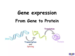
The Flow of Genetic Information • The information content of genes is in the specific sequences of nucleotides • The DNA inherited by an organism leads tospecific traits by dictating the synthesis of proteins • Proteins are the links between genotype and phenotype • Gene expression, the process by which DNA directs protein synthesis, includes two stages: transcription and translation
Evidence from the Study of Metabolic Defects • In 1902, British physician Archibald Garrod first suggested that genes dictate phenotypes through enzymes that catalyze specific chemical reactions • He thought symptoms of an inherited disease reflect an inability to synthesize a certain enzyme • Cells synthesize and degrade molecules in a series of steps, a metabolic pathway
Nutritional Mutants in Neurospora: Scientific Inquiry • George Beadle and Edward Tatum exposed bread mold to X-rays, creating mutants that were unable to survive on minimal media • Using crosses, they and their coworkers identified three classes of arginine-deficient mutants, each lacking a different enzyme necessary for synthesizing arginine • They developed a one gene–one enzyme hypothesis, which states that each gene dictates production of a specific enzyme
The Products of Gene Expression:A Developing Story • Some proteins aren’t enzymes, so researchers later revised the hypothesis: one gene–one protein • Many proteins are composed of several polypeptides, each of which has its own gene • Therefore, Beadle and Tatum’s hypothesis is now restated as the one gene–one polypeptide hypothesis • It is common to refer to gene products as proteins rather than polypeptides
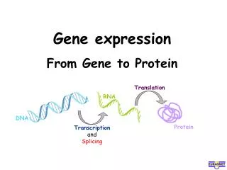
Basic Principles of Transcription and Translation • RNA is the bridge between genes and the proteins for which they code • Transcription is the synthesis of RNA using information in DNA • Transcription produces messenger RNA (mRNA) • Translation is the synthesis of a polypeptide, using information in the mRNA • Ribosomes are the sites of translation
In prokaryotes, translation of mRNA can begin before transcription has finished • In a eukaryotic cell, the nuclear envelope separates transcription from translation • Eukaryotic RNA transcripts are modified through RNA processing to yield the finished mRNA
Figure 17.3 Nuclear envelope DNA TRANSCRIPTION Pre-mRNA RNA PROCESSING mRNA NUCLEUS DNA TRANSCRIPTION CYTOPLASM CYTOPLASM mRNA TRANSLATION Ribosome Ribosome TRANSLATION Polypeptide Polypeptide (b) Eukaryotic cell (a) Bacterial cell
A primary transcript is the initial RNA transcript from any gene prior to processing • The central dogma is the concept that cells are governed by a cellular chain of command: DNA → RNA → protein
Figure 17.UN01 DNA RNA Protein
The Genetic Code • How are the instructions for assembling amino acids into proteins encoded into DNA? • There are 20 amino acids, but there are only four nucleotide bases in DNA • How many nucleotides correspond to anamino acid?
Codons: Triplets of Nucleotides • The flow of information from gene to protein is based on a triplet code: a series of nonoverlapping, three-nucleotide words • The words of a gene are transcribed into complementary nonoverlapping three-nucleotide words of mRNA • These words are then translated into a chain of amino acids, forming a polypeptide
Figure 17.4 DNA template strand 5′ 3′ C C C C G G T A A A A A T G G T T T G G C T C A 3′ 5′ TRANSCRIPTION U G G U U U G G C U C A mRNA 5′ 3′ Codon TRANSLATION Protein Gly Trp Phe Ser Amino acid
During transcription, one of the two DNA strands, called the template strand, provides a template for ordering the sequence of complementary nucleotides in an RNA transcript • The template strand is always the same strandfor a given gene
During translation, the mRNA base triplets, called codons, are read in the 5′ → 3′ direction • Each codon specifies the amino acid (one of 20)to be placed at the corresponding position alonga polypeptide
Cracking the Code • All 64 codons were deciphered by the mid-1960s • Of the 64 triplets, 61 code for amino acids; 3 triplets are “stop” signals to end translation • The genetic code is redundant (more than one codon may specify a particular amino acid) butnot ambiguous; no codon specifies more thanone amino acid • Codons must be read in the correct reading frame (correct groupings) in order for the specified polypeptide to be produced
Figure 17.5 Second mRNA base U C A G UAU UGU UUU UCU U Phe Tyr Cys UCC C UUC UAC UGC U Ser UUA UGA A UCA UAA Stop Stop Leu Trp UCG UAG Stop UGG G UUG CUU CGU CCU CAU U His CUC CCC CGC CAC C C Leu Pro Arg CUA CGA CCA CAA A Gln CAG CUG CCG CGG G First mRNA base (5′ end of codon) Third mRNA base (3′ end of codon) AUU AAU U ACU AGU Ser Asn Ile AUC ACC AGC C AAC A Thr AUA ACA AAA AGA A Lys Arg Met or start ACG AAG AGG AUG G GUU GCU U GAU GGU Asp GCC GAC GUC GGC C G Gly Val Ala GUA GCA GAA GGA A Glu GUG GCG GAG GGG G
Evolution of the Genetic Code • The genetic code is nearly universal, shared by the simplest bacteria to the most complex animals • Genes can be transcribed and translated after being transplanted from one species to another
Figure 17.6 (a) Tobacco plant expressing a firefly gene (b) Pig expressing a jellyfish gene
Concept 17.2: Transcription is the DNA-directed synthesis of RNA: A closer look • Transcription is the first stage of gene expression
Molecular Components of Transcription • RNA synthesis is catalyzed by RNA polymerase, which pries the DNA strands apart and joins together the RNA nucleotides • The RNA is complementary to the DNA template strand • RNA polymerase does not need any primer • RNA synthesis follows the same base-pairing rules as DNA, except that uracil substitutes for thymine
Figure 17.7-3 Promoter Transcription unit 3′ 5′ 3′ 5′ Start point RNA polymerase Initiation 1 3′ 5′ 3′ 5′ Template strand of DNA RNA transcript Unwound DNA Elongation 2 Rewound DNA 3′ 5′ 3′ 5′ 3′ 5′ Direction of transcription (“downstream”) RNA transcript Termination 3 3′ 5′ 5′ 3′ 3′ 5′ Completed RNA transcript
The DNA sequence where RNA polymerase attaches is called the promoter; in bacteria, the sequence signaling the end of transcription is called the terminator • The stretch of DNA that is transcribed is called a transcription unit
Synthesis of an RNA Transcript • The three stages of transcription • Initiation • Elongation • Termination
RNA Polymerase Binding and Initiation of Transcription • Promoters signal the transcriptional start point and usually extend several dozen nucleotide pairs upstream of the start point • Transcription factors mediate the binding of RNA polymerase and the initiation of transcription • The completed assembly of transcription factorsand RNA polymerase II bound to a promoter is called a transcription initiation complex • A promoter called a TATA box is crucial in forming the initiation complex in eukaryotes
Figure 17.8 Promoter Nontemplate strand DNA 5′ 3′ A A A T A T A A eukaryotic promoter 1 3′ 5′ A T A T T T T TATA box Start point Template strand Transcription factors 3′ 5′ Several transcription factors bind to DNA. 2 5′ 3′ RNA polymerase II Transcription factors 5′ 3′ Transcription initiation complex forms. 3′ 3 5′ 3′ 5′ RNA transcript Transcription initiation complex
Elongation of the RNA Strand • As RNA polymerase moves along the DNA, it untwists the double helix, 10 to 20 bases at a time • Transcription progresses at a rate of 40 nucleotides per second in eukaryotes • A gene can be transcribed simultaneously by several RNA polymerases • Nucleotides are added to the 3′ end of thegrowing RNA molecule
Figure 17.9 Nontemplate strand of DNA RNA nucleotides RNA polymerase C C T A A A T U 3′ 5′ C T 3′ end T G U A A G C C A C U A C C A 3′ 5′ A T A T G G T 5′ Direction of transcription Template strand of DNA Newly made RNA
Termination of Transcription • The mechanisms of termination are different in bacteria and eukaryotes • In bacteria, the polymerase stops transcription at the end of the terminator and the mRNA can be translated without further modification • In eukaryotes, RNA polymerase II transcribes the polyadenylation signal sequence; the RNA transcript is released 10–35 nucleotides past this polyadenylation sequence
Concept 17.3: Eukaryotic cells modify RNA after transcription • Enzymes in the eukaryotic nucleus modify pre-mRNA (RNA processing) before the genetic messages are dispatched to the cytoplasm • During RNA processing, both ends of the primary transcript are usually altered • Also, usually certain interior sections of the molecule are cut out, and the remaining parts spliced together
Alteration of mRNA Ends • Each end of a pre-mRNA molecule is modified in a particular way • The 5′ end receives a modified nucleotide 5′ cap • The 3′ end gets a poly-A tail • These modifications share several functions • They seem to facilitate the export of mRNA to the cytoplasm • They protect mRNA from hydrolytic enzymes • They help ribosomes attach to the 5′ end
Figure 17.10 50–250 adenine nucleotides added to the 3′ end A modified guanine nucleotide added to the 5′ end Polyadenylation signal Region that includes protein-coding segments 3′ 5′ … G P P P AAUAAA AAA AAA Start codon Stop codon 5′ Cap 5′ UTR 3′ UTR Poly-A tail
Split Genes and RNA Splicing • Most eukaryotic genes and their RNA transcripts have long noncoding stretches of nucleotides that lie between coding regions • These noncoding regions are called intervening sequences, or introns • The other regions are called exons because they are eventually expressed, usually translated into amino acid sequences • RNA splicing removes introns and joins exons, creating an mRNA molecule with a continuous coding sequence
Figure 17.11 Pre-mRNA Intron Intron 5′ Cap Poly-A tail Introns cut out and exons spliced together 1–30 31–104 105–146 mRNA 5′ Cap Poly-A tail 1–146 5′ UTR 3′ UTR Coding segment
In some cases, RNA splicing is carried out by spliceosomes • Spliceosomes consist of a variety of proteins and several small nuclear ribonucleoproteins (snRNPs) that recognize the splice sites • The RNAs of the spliceosome also catalyze the splicing reaction
Figure 17.12 Small RNAs Spliceosome 5′ Pre-mRNA Exon 2 Exon 1 Intron Spliceosome components mRNA Cut-out intron 5′ Exon 1 Exon 2
Ribozymes • Ribozymes are catalytic RNA molecules that function as enzymes and can splice RNA • The discovery of ribozymes rendered obsolete the belief that all biological catalysts were proteins
Three properties of RNA enable it to function asan enzyme • It can form a three-dimensional structure because of its ability to base-pair with itself • Some bases in RNA contain functional groupsthat may participate in catalysis • RNA may hydrogen-bond with other nucleicacid molecules
The Functional and Evolutionary Importance of Introns • Some introns contain sequences that may regulate gene expression • Some genes can encode more than one kind of polypeptide, depending on which segments are treated as exons during splicing • This is called alternative RNA splicing • Consequently, the number of different proteinsan organism can produce is much greater thanits number of genes
Proteins often have a modular architecture consisting of discrete regions called domains • In many cases, different exons code for the different domains in a protein • Exon shuffling may result in the evolution of new proteins
Figure 17.13 Gene DNA Exon 1 Intron Exon 2 Intron Exon 3 Transcription RNA processing Translation Domain 3 Domain 2 Domain 1 Polypeptide
Concept 17.4: Translation is the RNA-directed synthesis of a polypeptide: A closer look • Genetic information flows from mRNA to protein through the process of translation
Molecular Components of Translation • A cell translates an mRNA message into protein with the help of transfer RNA (tRNA) • tRNAs transfer amino acids to the growing polypeptide in a ribosome • Translation is a complex process in terms of its biochemistry and mechanics
Figure 17.14 Amino acids Polypeptide tRNA with amino acid attached Ribosome Trp Gly Phe tRNA C C C Anticodon C G A A A A U G G U U U G G C Codons 5′ 3′ mRNA
The Structure and Function of Transfer RNA • Molecules of tRNA are not identical • Each carries a specific amino acid on one end • Each has an anticodon on the other end; the anticodon base-pairs with a complementary codon on mRNA
A tRNA molecule consists of a single RNA strand that is only about 80 nucleotides long • Flattened into one plane to reveal its base pairing, a tRNA molecule looks like a cloverleaf
Because of hydrogen bonds, tRNA actually twists and folds into a three-dimensional molecule • tRNA is roughly L-shaped
Figure 17.15 3′ Amino acid attachment site A C C 5′ A Amino acid attachment site C G C G C G 5′ U G 3′ U A U A A U U C G * G A U * C A A C A C U * C G * U U G G G * Hydrogen bonds G A G C C * * C G A G U * * A G C G Hydrogen bonds G C A U * G * A A C A A G * U 3′ 5′ A G A Anticodon Anticodon Anticodon (a) Two-dimensional structure (b) (c) Symbol used in this book Three-dimensional structure
Accurate translation requires two steps • First: a correct match between a tRNA and an amino acid, done by the enzyme aminoacyl-tRNAsynthetase • Second: a correct match between the tRNA anticodon and an mRNA codon • Flexible pairing at the third base of a codon is called wobble and allows some tRNAs to bind to more than one codon
Figure 17.16-3 Tyrosine (Tyr) (amino acid) 1 Amino acid and tRNA enter active site. Tyrosyl-tRNA synthetase Tyr-tRNA Aminoacyl-tRNA synthetase U ATP A A AMP + 2 P i tRNA Complementary tRNA anticodon Amino acid 3 Using ATP, synthetase catalyzes covalent bonding. Aminoacyl tRNA released. 2 Computer model