Download
1 / 24
250 likes | 306 Views
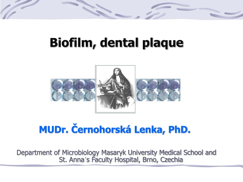
Biofilm, dental plaque. MUDr. Černohorská Lenka, PhD. Department of Microbiology Masaryk University Medical School and St. Anna´s Faculty Hospital, Brno, Czechia. M icrobial growth. P lanktonic form M icrobial cells float freely in a fluid B iofilm form

E N D
Biofilm, dental plaque MUDr. Černohorská Lenka, PhD. Department of Microbiology Masaryk University Medical School and St. Anna´s Faculty Hospital, Brno, Czechia
Microbial growth • Planktonic form Microbial cells float freely in a fluid • Biofilm form Microbial cells stick to one another and to a solid surface and form a community connected by an extracellular matter
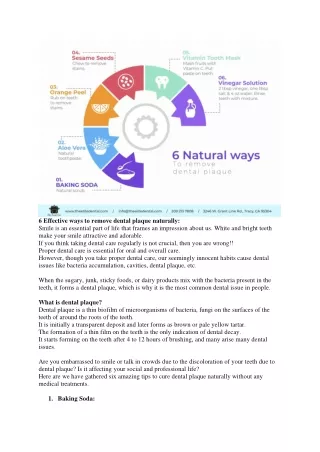
Examples of biofilm • Have you ever slipped on a wet stone in a creek?It was biofilm that you slipped on • Have you an aquarium and do you clean its walls?If you do, what you wipe from them is the biofilm formed by algae • Do you clean your teeth regularly? I hope so and by doing this you remove the biofilm called dental plaque
Definition of biofilm Microbial biofilm is a 3D strucuture which: • forms at the boundary of phases (usually of the solid and fluid phase) • is surrounded by an extracellular matter, in which a complex system of channels forms
Stages of biofilm development • Direct contact of aplanktonicbacteriawith asurface+ • Attachment to this surface • Adhesion, growth, andaggregationof cells into microcolonies • Production of polymeric matrix • Formation of three-dimensional structure known as biofilm
Development of biofilm 1. Attraction – in mobile bacteria due to flagella 2. Adhesion – via bacterial adhesins fimbriae (pilli):colonization factors of enteropathogenic E. coli proteins and lipopolysaccharides of outer membrane (G-negative bacteria) slime (coagulase-negative + S. aureus) curli (E. coli)
Development of biofilm – 3. Aggregation • Movement - by flagella (E. coli), by means of fimbriae (P.aeruginosa), convergent – aggregates of different species (coaggregation of Str. gordonii + F. nucleatum in dental plaque) • Multiplication - aggregation + cell division in aggregates lead to the development of microcolonies • Quorum sensing- during division individual cells emit chemical signals (homoserinlactones in P. aeruginosa). After reaching a particular number of cells (quorum) the elevated concentration of signals causes the change of cellular properties: switching off some genes, expression of other genes, production of new molecules
Development of biofilm 4. Accumulation: production of exopolysaccharidesleads to the development of typical biofilm architecture - colanic acid (E. coli), alginate (P. aeruginosa), polysaccharide intercellular adhesin (S. epidermidis) 5. Dispersal: after reaching the critical amount of biomass + after the reduction of nutrients in the environment the character of surface cells changes (for. ex. P. aeruginosathe superficial cells produce lyase and flagellin, superficial layer of biofilm starts to disintegrate, cells grow flagella and get loose of biofilm) The cells drift away as a planktonic population to look for more suitable environment and tocolonize new surfaces. The cycle closes…
Architecture of biofilm Candida albicans biofilm. Toluidin blue -mushroom-like structure of the biofilm Alcian blue has coloured extracellular polysaccharides. Photo: Veronika Holá
Architecture of biofilm Depends on the concentration of nutrients • <10 mg/L (mountain streams, lakes, open sea) heterogeneous mosaic - a thin layer + columns of microcolonies • 10-1000 mg/L (majority of our rivers and ponds) complex system with channels (created by mushroom-like, partially merging microcolonies) • 1000 mg/L (in the environment of macroorganism) compact biofilm (almost without traces of channels)
Main importanceof biofilm formation Bacteriaharbored inside are protected against: • antibiotic action • host´s immune response • disinfection
Researching methods + - • In bacterial strains(S. aureus, E. coli, P. aeruginosa etc.) biofilm can be detected by themodified Christensen method • Biofilm susceptibility testing:MBIC (minumum biofilm inhibitory concentration) was determined • MBIC was compared with MIC • Synergy testing:FBIC (fractionate biofilm inhibitory concentration) was calculated as follows: FBICs (∑FBIC) = MBIC ATB A in combination+ MBIC ATB Bin combination MBIC ATB A alone MBIC ATB B alone Combinations of antimicrobial agents: synergistic (∑FBIC ≤ 0,5)partially synergistic (∑FBIC > 0.5 a ≤1) indifferent (∑FBIC > 1 a ≤ 4)antagonistic (∑FBIC > 4)
Antibiotic susceptibility of staphylococci isolates Planktonic bacteria (MIC) Biofilm-forming bacteria (MBIC) Abbreviations:ams - ampicillin/sulbactam, ch - chloramphenicol, ery - erytromycin, te - tetracyclin, cli - clindamycin, tei - teicoplanin, van - vancomycin, ofl - ofloxacin
The inefficiency of antibiotics may be due to: • Polyanionic charge of sessile cells • Decreased bacterialgrowth • Diffusion barrier of glycocalyx • Reactionwithbiofilm matrix • Formation of protectedphenotypes • Mechanism ofintercellular signalling • Host´s immune response mechanisms…
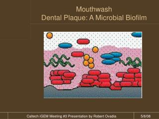
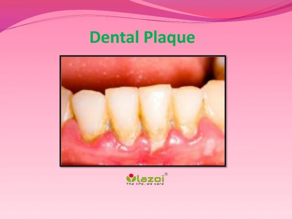
Dental plaque • Sticky microbial layer on the teeth surface, composed oflivinganddead bacteriaand their products +components of host originoriginated from salivas • Cannotbewashed, is removable only mechanically • Supragingival andsubgingival • Differsin morphology and microbialcomponents • Subgingival plaque is divided intoadherent and non-adherent • Composition of plaquedepends onitsage and localization, speed of plaque formationis individually • Contains many bacterial species
Plaque development • On the teeth surface forms thinlayer of saliva glycoproteins = pelicula. • The salivastransportoral bacteria to this surface • First adhereG+cocci androdsdue to surface adhesins. ToG+ bacteria adhere the other • Aggregated bacteriadivides andform microcolonies • Productionof exopolysaccharides
Metabolism of bacteriachangestheirenvironment and enable plaque formation alsotoanother microbial species • Presence of saccharosis accelerates mature of plaque • Bacteria free themselves from the outer layers, innerlayersformdentalstone • pH under 5,5 -demineralisationof enamel andformation of dental caries
Reaction of gingiva • Exsudate formation • Inflammation – gingivitis - damages function ofjoining epitelium andplaque penetratesto subgingival area • Older and stronger plaque – more symptoms Kolenbrander et al., 2002
Subgingival plaque 2types: adherent andnon-adherent • Adherent plaque - sessileon thestub = G+rods andfibres (actinomycets) andG+cocci • Non-adherentplaque - between adherent plaque andsurfaceof softgingival tissue = G-mobile anaerobes
Non-adherent plaque MobileG- anaerobes: • Porphyromonades (P. gingivalis), • Prevotela (P. nigrescens) • Fusobacterium (F. nucleatum subsp. polymorphum) • Treponema (T. denticola). More patogennousthanG+ cocci androds. Gingivitisbecame worse = development ofparodontal snout.
Plaque onthe teeth implantates • Variousbacteria • Group of Streptococcusmutans andsanguinis, yeast (Candida) - oncontact area with mucous • Anaerobes:G+rods+ Actinomyces israelii+ veillonela • Staphylococci (S. aureus)
Teethstone • 80% minerals, mainlyhydroxylapatit, lesscalcium carbonate andmagnesium phosphateand organic substances (rest of microbial cells, epitelia and mucin) • Stoneabove gingivacontainsmainly G+bacteria, subgingival stoneG -bacteria • Structure - porosity and roughsurfaceof dentalstone - filamentous bacteriain plaqueare orientedvertical+palisade tosurface of the teeth - storageofmicrobial components toxic for parodontic tissue